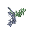
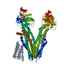
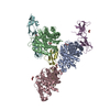
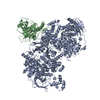
Entry Database : PDB / ID : 4yocTitle Crystal Structure of human DNMT1 and USP7/HAUSP complex DNA (cytosine-5)-methyltransferase 1 Ubiquitin carboxyl-terminal hydrolase 7 Keywords / / / / / Function / homology Function Domain/homology Component
/ / / / / / / / / / / / / / / / / / / / / / / / / / / / / / / / / / / / / / / / / / / / / / / / / / / / / / / / / / / / / / / / / / / / / / / / / / / / / / / / / / / / / / / / / / / / / / / / / / / / / / / / / / / / / / / / / / / / / / / / / / / / / / / / / / / / / / / / / / Biological species Homo sapiens (human)Method / / / / Resolution : 2.916 Å Authors Cheng, J. / Yang, H. / Fang, J. / Gong, R. / Wang, P. / Li, Z. / Xu, Y. Journal : Nat Commun / Year : 2015Title : Molecular mechanism for USP7-mediated DNMT1 stabilization by acetylation.Authors : Cheng, J. / Yang, H. / Fang, J. / Ma, L. / Gong, R. / Wang, P. / Li, Z. / Xu, Y. History Deposition Mar 11, 2015 Deposition site / Processing site Revision 1.0 May 27, 2015 Provider / Type Revision 1.1 Nov 8, 2023 Group Data collection / Database references ... Data collection / Database references / Derived calculations / Refinement description / Source and taxonomy Category chem_comp_atom / chem_comp_bond ... chem_comp_atom / chem_comp_bond / database_2 / entity_src_gen / pdbx_initial_refinement_model / pdbx_struct_oper_list Item _database_2.pdbx_DOI / _database_2.pdbx_database_accession ... _database_2.pdbx_DOI / _database_2.pdbx_database_accession / _entity_src_gen.pdbx_alt_source_flag / _pdbx_struct_oper_list.symmetry_operation
Show all Show less
 Movie
Movie Controller


 Open data
 Basic information
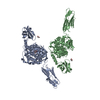
 Components
 Keywords
 Function and homology information
 Homo sapiens (human)
 X-RAY DIFFRACTION /
  Authors
 Citation
 Structure visualization
 Downloads & links
 Other downloads
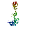


 PDBj
PDBj


 Assembly
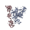


 Mass: 65.409 Da / Num. of mol.: 2 / Source method: obtained synthetically / Formula: Zn
 Mass: 18.015 Da / Num. of mol.: 11 / Source method: isolated from a natural source / Formula: H2O
 Sample preparation
 / Beamline: BL17U / Wavelength: 1 Å
 Processing