 Movie
Movie Controller


+ Open data

- Basic information
| Entry | Database: PDB / ID: 5nvs | |||||||||
|---|---|---|---|---|---|---|---|---|---|---|
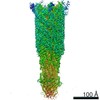
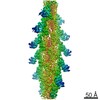
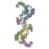
| Title | Human cytoplasmic dynein-1 tail in the twisted N-terminus state | |||||||||
 Components Components |
| |||||||||
 Keywords Keywords | MOTOR PROTEIN / dynein / tail complex | |||||||||
| Biological species |  Homo sapiens (human) Homo sapiens (human) | |||||||||
| Method | ELECTRON MICROSCOPY / single particle reconstruction / cryo EM / Resolution: 8.4 Å | |||||||||
 Authors Authors | Zhang, K. / Foster, H.E. / Carter, A.P. | |||||||||
| Funding support |  United Kingdom, 2items United Kingdom, 2items
| |||||||||
 Citation Citation | Journal: Cell / Year: 2017 Title: Cryo-EM Reveals How Human Cytoplasmic Dynein Is Auto-inhibited and Activated. Authors: Kai Zhang / Helen E Foster / Arnaud Rondelet / Samuel E Lacey / Nadia Bahi-Buisson / Alexander W Bird / Andrew P Carter /   Abstract: Cytoplasmic dynein-1 binds dynactin and cargo adaptor proteins to form a transport machine capable of long-distance processive movement along microtubules. However, it is unclear why dynein-1 moves ...Cytoplasmic dynein-1 binds dynactin and cargo adaptor proteins to form a transport machine capable of long-distance processive movement along microtubules. However, it is unclear why dynein-1 moves poorly on its own or how it is activated by dynactin. Here, we present a cryoelectron microscopy structure of the complete 1.4-megadalton human dynein-1 complex in an inhibited state known as the phi-particle. We reveal the 3D structure of the cargo binding dynein tail and show how self-dimerization of the motor domains locks them in a conformation with low microtubule affinity. Disrupting motor dimerization with structure-based mutagenesis drives dynein-1 into an open form with higher affinity for both microtubules and dynactin. We find the open form is also inhibited for movement and that dynactin relieves this by reorienting the motor domains to interact correctly with microtubules. Our model explains how dynactin binding to the dynein-1 tail directly stimulates its motor activity. | |||||||||
| History |
|
- Structure visualization
| Movie |
 Movie viewer Movie viewer |
|---|---|
| Structure viewer | Molecule: MolmilJmol/JSmol |
- Downloads & links
-Download
| PDBx/mmCIF format | 5nvs.cif.gz | 445.8 KB | Display | PDBx/mmCIF format |
|---|---|---|---|---|
| PDB format | pdb5nvs.ent.gz | 353 KB | Display | PDB format |
| PDBx/mmJSON format | 5nvs.json.gz | Tree view | PDBx/mmJSON format | |
| Others |  Other downloads Other downloads |
-Validation report
| Arichive directory | https://data.pdbj.org/pub/pdb/validation_reports/nv/5nvsftp://data.pdbj.org/pub/pdb/validation_reports/nv/5nvs | HTTPS FTP |
|---|
-Related structure data
| Related structure data | 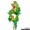 3703MC 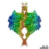 3698C  3704C 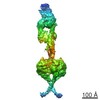 3705C 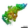 3706C 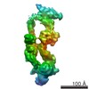 3707C  5nugC  5nvuC  5nw4C C: citing same article ( M: map data used to model this data |
|---|---|
| Similar structure data |


-Links
 PDBj
PDBj
- Assembly
| Deposited unit | 
|
|---|---|
| 1 |
|
-Components
-Dynein heavy ... , 2 types, 2 molecules AB
| #1: Protein | Mass: 79335.086 Da / Num. of mol.: 1 Source method: isolated from a genetically manipulated source Source: (gene. exp.) Homo sapiens (human) / Production host:   Spodoptera frugiperda (fall armyworm) Spodoptera frugiperda (fall armyworm) |
|---|---|
| #3: Protein | Mass: 76016.109 Da / Num. of mol.: 1 Source method: isolated from a genetically manipulated source Source: (gene. exp.) Homo sapiens (human) / Production host: Spodoptera frugiperda (fall armyworm) |
-Protein , 5 types, 8 molecules DCIJKLRS
| #2: Protein | Mass: 29804.609 Da / Num. of mol.: 2 Source method: isolated from a genetically manipulated source Source: (gene. exp.) Homo sapiens (human) / Production host: Spodoptera frugiperda (fall armyworm)#8: Protein | Mass: 7251.930 Da / Num. of mol.: 2 Source method: isolated from a genetically manipulated source Source: (gene. exp.) Homo sapiens (human) / Production host: Spodoptera frugiperda (fall armyworm)#9: Protein | | Mass: 8783.818 Da / Num. of mol.: 1 Source method: isolated from a genetically manipulated source Source: (gene. exp.) Homo sapiens (human) / Production host: Spodoptera frugiperda (fall armyworm)#10: Protein | | Mass: 8868.924 Da / Num. of mol.: 1 Source method: isolated from a genetically manipulated source Source: (gene. exp.) Homo sapiens (human) / Production host: Spodoptera frugiperda (fall armyworm)#13: Protein | Mass: 10230.603 Da / Num. of mol.: 2 Source method: isolated from a genetically manipulated source Source: (gene. exp.) Homo sapiens (human) / Production host: Spodoptera frugiperda (fall armyworm) |
|---|
-Dynein light intermediate ... , 2 types, 2 molecules FE
| #4: Protein | Mass: 25379.143 Da / Num. of mol.: 1 Source method: isolated from a genetically manipulated source Source: (gene. exp.) Homo sapiens (human) / Production host: Spodoptera frugiperda (fall armyworm) |
|---|---|
| #5: Protein | Mass: 25123.830 Da / Num. of mol.: 1 Source method: isolated from a genetically manipulated source Source: (gene. exp.) Homo sapiens (human) / Production host: Spodoptera frugiperda (fall armyworm) |
-N-terminal dimerization ... , 2 types, 2 molecules 21
| #6: Protein | Mass: 10656.127 Da / Num. of mol.: 1 Source method: isolated from a genetically manipulated source Source: (gene. exp.) Homo sapiens (human) / Production host: Spodoptera frugiperda (fall armyworm) |
|---|---|
| #7: Protein | Mass: 10571.022 Da / Num. of mol.: 1 Source method: isolated from a genetically manipulated source Source: (gene. exp.) Homo sapiens (human) / Production host: Spodoptera frugiperda (fall armyworm) |
-Intermediate ... , 2 types, 2 molecules MN
| #11: Protein/peptide | Mass: 2315.846 Da / Num. of mol.: 1 Source method: isolated from a genetically manipulated source Source: (gene. exp.) Homo sapiens (human) / Production host: Spodoptera frugiperda (fall armyworm) |
|---|---|
| #12: Protein/peptide | Mass: 2486.056 Da / Num. of mol.: 1 Source method: isolated from a genetically manipulated source Source: (gene. exp.) Homo sapiens (human) / Production host: Spodoptera frugiperda (fall armyworm) |
-Experimental details
-Experiment
| Experiment | Method: ELECTRON MICROSCOPY |
|---|---|
| EM experiment | Aggregation state: PARTICLE / 3D reconstruction method: single particle reconstruction |
- Sample preparation
| Component | Name: Human cytoplasmic dynein-1 tail in the twisted N-terminus state Type: COMPLEX / Entity ID: all / Source: RECOMBINANT |
|---|---|
| Molecular weight | Value: 1.4 MDa / Experimental value: YES |
| Source (natural) | Organism: Homo sapiens (human) |
| Source (recombinant) | Organism: Spodoptera frugiperda (fall armyworm) |
| Buffer solution | pH: 7.4 |
| Specimen | Conc.: 0.15 mg/ml / Embedding applied: NO / Shadowing applied: NO / Staining applied: NO / Vitrification applied: YES |
| Vitrification | Cryogen name: ETHANE |
- Electron microscopy imaging
| Experimental equipment |  Model: Titan Krios / Image courtesy: FEI Company |
|---|---|
| Microscopy | Model: FEI TITAN KRIOS |
| Electron gun | Electron source:  FIELD EMISSION GUN / Accelerating voltage: 300 kV / Illumination mode: FLOOD BEAM FIELD EMISSION GUN / Accelerating voltage: 300 kV / Illumination mode: FLOOD BEAM |
| Electron lens | Mode: BRIGHT FIELD |
| Image recording | Electron dose: 1.6 e/Å2 / Film or detector model: FEI FALCON II (4k x 4k) |
- Processing
| EM software |
| ||||||||||||||||||||
|---|---|---|---|---|---|---|---|---|---|---|---|---|---|---|---|---|---|---|---|---|---|
| CTF correction | Type: PHASE FLIPPING AND AMPLITUDE CORRECTION | ||||||||||||||||||||
| Particle selection | Details: Particles were selected on the phase-flipped micrographs by Gautomatch. | ||||||||||||||||||||
| Symmetry | Point symmetry: C1 (asymmetric) | ||||||||||||||||||||
| 3D reconstruction | Resolution: 8.4 Å / Resolution method: FSC 0.143 CUT-OFF / Num. of particles: 187830 / Symmetry type: POINT |
