Movie
Movie Controller

[English] 日本語
 Yorodumi
- PDB-5k0z: Cryo-EM structure of lactate dehydrogenase (LDH) in inhibitor-bou... -
+ Open data

- Basic information
| Entry | Database: PDB / ID: 5k0z | ||||||
|---|---|---|---|---|---|---|---|
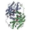
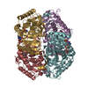
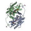
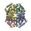
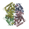
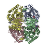
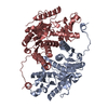
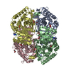
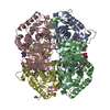
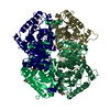
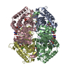
| Title | Cryo-EM structure of lactate dehydrogenase (LDH) in inhibitor-bound state | ||||||
 Components Components | L-lactate dehydrogenase B chain | ||||||
 Keywords Keywords | OXIDOREDUCTASE / lactate dehydrogenase / small metabolic complex / small molecule inhibitor | ||||||
| Function / homology |  Function and homology information Function and homology informationPyruvate metabolism / Pyruvate metabolism / L-lactate dehydrogenase / oxidoreductase complex / pyruvate metabolic process / L-lactate dehydrogenase (NAD+) activity / lactate metabolic process / mitochondrial inner membrane / membrane raft / mitochondrion ...Pyruvate metabolism / Pyruvate metabolism / L-lactate dehydrogenase / oxidoreductase complex / pyruvate metabolic process / L-lactate dehydrogenase (NAD+) activity / lactate metabolic process / mitochondrial inner membrane / membrane raft / mitochondrion / identical protein binding / cytosol Similarity search - Function | ||||||
| Biological species |  | ||||||
| Method | ELECTRON MICROSCOPY / single particle reconstruction / cryo EM / Resolution: 2.8 Å | ||||||
 Authors Authors | Merk, A. / Bartesaghi, A. / Banerjee, S. / Falconieri, V. / Rao, P. / Earl, L. / Milne, J. / Subramaniam, S. | ||||||
 Citation Citation | Journal: Cell / Year: 2016 Title: Breaking Cryo-EM Resolution Barriers to Facilitate Drug Discovery. Authors: Alan Merk / Alberto Bartesaghi / Soojay Banerjee / Veronica Falconieri / Prashant Rao / Mindy I Davis / Rajan Pragani / Matthew B Boxer / Lesley A Earl / Jacqueline L S Milne / Sriram Subramaniam /  Abstract: Recent advances in single-particle cryoelecton microscopy (cryo-EM) are enabling generation of numerous near-atomic resolution structures for well-ordered protein complexes with sizes ≥ ∼200 kDa. ...Recent advances in single-particle cryoelecton microscopy (cryo-EM) are enabling generation of numerous near-atomic resolution structures for well-ordered protein complexes with sizes ≥ ∼200 kDa. Whether cryo-EM methods are equally useful for high-resolution structural analysis of smaller, dynamic protein complexes such as those involved in cellular metabolism remains an important question. Here, we present 3.8 Å resolution cryo-EM structures of the cancer target isocitrate dehydrogenase (93 kDa) and identify the nature of conformational changes induced by binding of the allosteric small-molecule inhibitor ML309. We also report 2.8-Å- and 1.8-Å-resolution structures of lactate dehydrogenase (145 kDa) and glutamate dehydrogenase (334 kDa), respectively. With these results, two perceived barriers in single-particle cryo-EM are overcome: (1) crossing 2 Å resolution and (2) obtaining structures of proteins with sizes < 100 kDa, demonstrating that cryo-EM can be used to investigate a broad spectrum of drug-target interactions and dynamic conformational states. | ||||||
| History |
|
- Structure visualization
| Movie |
Movie viewer |
|---|---|
| Structure viewer | Molecule: MolmilJmol/JSmol |
- Downloads & links
-Download
| PDBx/mmCIF format | 5k0z.cif.gz | 238 KB | Display | PDBx/mmCIF format |
|---|---|---|---|---|
| PDB format | pdb5k0z.ent.gz | 195.5 KB | Display | PDB format |
| PDBx/mmJSON format | 5k0z.json.gz | Tree view | PDBx/mmJSON format | |
| Others |  Other downloads Other downloads |
-Validation report
| Arichive directory | https://data.pdbj.org/pub/pdb/validation_reports/k0/5k0zftp://data.pdbj.org/pub/pdb/validation_reports/k0/5k0z | HTTPS FTP |
|---|
-Related structure data
| Related structure data | 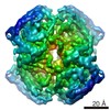 8191MC 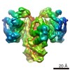 8192C 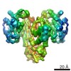 8193C 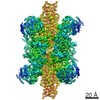 8194C 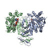 5k10C 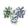 5k11C 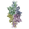 5k12C M: map data used to model this data C: citing same article ( |
|---|---|
| Similar structure data |
-Links
 PDBj
PDBj


- Assembly
| Deposited unit | 
|
|---|---|
| 1 |
|
-Components
| #1: Protein | Mass: 36115.656 Da / Num. of mol.: 4 Source method: isolated from a genetically manipulated source Source: (gene. exp.) #2: Water | ChemComp-HOH / |  Mass: 18.015 Da / Num. of mol.: 41 / Source method: isolated from a natural source / Formula: H2O Mass: 18.015 Da / Num. of mol.: 41 / Source method: isolated from a natural source / Formula: H2O |
|---|
-Experimental details
-Experiment
| Experiment | Method: ELECTRON MICROSCOPY |
|---|---|
| EM experiment | Aggregation state: PARTICLE / 3D reconstruction method: single particle reconstruction |
- Sample preparation
| Component | Name: Lactate dehydrogenase (LDH) in complex with GSK2837808A Type: COMPLEX / Entity ID: #1 / Source: RECOMBINANT |
|---|---|
| Molecular weight | Value: 0.145 MDa / Experimental value: NO |
| Source (natural) | Organism: |
| Source (recombinant) | Organism: Bacteria (eubacteria) / Plasmid: unknown |
| Buffer solution | pH: 7.4 / Details: Phosphate-buffered saline |
| Buffer component | Name: PBS |
| Specimen | Conc.: 1.5 mg/ml / Embedding applied: NO / Shadowing applied: NO / Staining applied: NO / Vitrification applied: YES |
| Specimen support | Grid material: COPPER / Grid mesh size: 300 divisions/in. / Grid type: Quantifoil R1.2/1.3 |
| Vitrification | Instrument: LEICA EM GP / Cryogen name: ETHANE / Details: Plunged into liquid ethane (LEICE EM GP) |
- Electron microscopy imaging
| Experimental equipment |  Model: Titan Krios / Image courtesy: FEI Company |
|---|---|
| Microscopy | Model: FEI TITAN KRIOS |
| Electron gun | Electron source:  FIELD EMISSION GUN / Accelerating voltage: 300 kV / Illumination mode: FLOOD BEAM FIELD EMISSION GUN / Accelerating voltage: 300 kV / Illumination mode: FLOOD BEAM |
| Electron lens | Mode: BRIGHT FIELD / Nominal magnification: 270000 X / Calibrated magnification: 101000 X / Nominal defocus max: 2200 nm / Nominal defocus min: 800 nm / Cs: 2.7 mm |
| Specimen holder | Cryogen: NITROGEN / Specimen holder model: FEI TITAN KRIOS AUTOGRID HOLDER / Temperature (max): 79.8 K / Temperature (min): 79.6 K |
| Image recording | Average exposure time: 0.2 sec. / Electron dose: 60 e/Å2 / Detector mode: SUPER-RESOLUTION / Film or detector model: GATAN K2 QUANTUM (4k x 4k) / Num. of real images: 1707 |
| EM imaging optics | Energyfilter name: GIF Quantum / Energyfilter upper: 20 eV / Energyfilter lower: 0 eV |
| Image scans | Movie frames/image: 60 / Used frames/image: 0-29 |
- Processing
| EM software |
| ||||||||||||||||||||||||||||||||
|---|---|---|---|---|---|---|---|---|---|---|---|---|---|---|---|---|---|---|---|---|---|---|---|---|---|---|---|---|---|---|---|---|---|
| CTF correction | Type: PHASE FLIPPING AND AMPLITUDE CORRECTION | ||||||||||||||||||||||||||||||||
| Particle selection | Num. of particles selected: 508402 | ||||||||||||||||||||||||||||||||
| Symmetry | Point symmetry: D2 (2x2 fold dihedral) | ||||||||||||||||||||||||||||||||
| 3D reconstruction | Resolution: 2.8 Å / Resolution method: FSC 0.143 CUT-OFF / Num. of particles: 50865 / Algorithm: FOURIER SPACE Details: The primary map in this entry corresponds to the uncorrected reconstruction. A version sharpened using a B-factor of -150 is provided as additional volume data. Symmetry type: POINT | ||||||||||||||||||||||||||||||||
| Atomic model building | Protocol: FLEXIBLE FIT / Space: REAL | ||||||||||||||||||||||||||||||||
| Atomic model building | PDB-ID: 1I0Z Accession code: 1I0Z / Source name: PDB / Type: experimental model |