 Movie
Movie Controller

[English] 日本語
 Yorodumi
- EMDB-5559: Cryo-em map of one molecule of factor VIII light chain from helic... -
+ Open data

- Basic information
| Entry | Database: EMDB / ID: EMD-5559 | |||||||||
|---|---|---|---|---|---|---|---|---|---|---|
| Title | Cryo-em map of one molecule of factor VIII light chain from helically organized factor VIII light chain molecules bound to lipid nanotubes | |||||||||
 Map data Map data | Cropped volume from helical reconstruction of membrane-bound Factor vIII light chain bound to single bilayer lipid nanotubes (EMD-5540) with a Gaussian filter of 3.0 applied | |||||||||
 Sample Sample |
| |||||||||
 Keywords Keywords | membrane binding / factor VIII light chain / helical organization / cryo-EM | |||||||||
| Function / homology |  Function and homology information Function and homology informationDefective F8 accelerates dissociation of the A2 domain / Defective F8 binding to the cell membrane / Defective F8 secretion / Defective F8 sulfation at Y1699 / Gamma carboxylation, hypusinylation, hydroxylation, and arylsulfatase activation / Defective F8 binding to von Willebrand factor / blood coagulation, intrinsic pathway / Cargo concentration in the ER / Defective factor IX causes thrombophilia / Defective cofactor function of FVIIIa variant ...Defective F8 accelerates dissociation of the A2 domain / Defective F8 binding to the cell membrane / Defective F8 secretion / Defective F8 sulfation at Y1699 / Gamma carboxylation, hypusinylation, hydroxylation, and arylsulfatase activation / Defective F8 binding to von Willebrand factor / blood coagulation, intrinsic pathway / Cargo concentration in the ER / Defective factor IX causes thrombophilia / Defective cofactor function of FVIIIa variant / Defective F9 variant does not activate FX / COPII-mediated vesicle transport / COPII-coated ER to Golgi transport vesicle / Defective F8 cleavage by thrombin / Common Pathway of Fibrin Clot Formation / Intrinsic Pathway of Fibrin Clot Formation / endoplasmic reticulum-Golgi intermediate compartment membrane / platelet alpha granule lumen / acute-phase response / Golgi lumen / blood coagulation / Platelet degranulation / oxidoreductase activity / copper ion binding / endoplasmic reticulum lumen / extracellular space / extracellular region / plasma membrane Similarity search - Function | |||||||||
| Biological species |  Homo sapiens (human) Homo sapiens (human) | |||||||||
| Method | helical reconstruction / cryo EM / Resolution: 15.0 Å | |||||||||
 Authors Authors | Stoilova-McPhie S / Lynch GC / Ludtke S / Pettitt BM | |||||||||
 Citation Citation | Journal: Biopolymers / Year: 2013 Title: Domain organization of membrane-bound factor VIII. Authors: Svetla Stoilova-McPhie / Gillian C Lynch / Steven Ludtke / B Montgomery Pettitt /  Abstract: Factor VIII (FVIII) is the blood coagulation protein which when defective or deficient causes for hemophilia A, a severe hereditary bleeding disorder. Activated FVIII (FVIIIa) is the cofactor to the ...Factor VIII (FVIII) is the blood coagulation protein which when defective or deficient causes for hemophilia A, a severe hereditary bleeding disorder. Activated FVIII (FVIIIa) is the cofactor to the serine protease factor IXa (FIXa) within the membrane-bound Tenase complex, responsible for amplifying its proteolytic activity more than 100,000 times, necessary for normal clot formation. FVIII is composed of two noncovalently linked peptide chains: a light chain (LC) holding the membrane interaction sites and a heavy chain (HC) holding the main FIXa interaction sites. The interplay between the light and heavy chains (HCs) in the membrane-bound state is critical for the biological efficiency of FVIII. Here, we present our cryo-electron microscopy (EM) and structure analysis studies of human FVIII-LC, when helically assembled onto negatively charged single lipid bilayer nanotubes. The resolved FVIII-LC membrane-bound structure supports aspects of our previously proposed FVIII structure from membrane-bound two-dimensional (2D) crystals, such as only the C2 domain interacts directly with the membrane. The LC is oriented differently in the FVIII membrane-bound helical and 2D crystal structures based on EM data, and the existing X-ray structures. This flexibility of the FVIII-LC domain organization in different states is discussed in the light of the FVIIIa-FIXa complex assembly and function. | |||||||||
| History |
|
- Structure visualization
| Movie |
Movie viewer |
|---|---|
| Structure viewer | EM map: SurfViewMolmilJmol/JSmol |
| Supplemental images |

- Downloads & links
-EMDB archive
| Map data | emd_5559.map.gz | 289.1 KB | EMDB map data format | |
|---|---|---|---|---|
| Header (meta data) | emd-5559-v30.xmlemd-5559.xml | 12.2 KB 12.2 KB | Display Display | EMDB header |
| Images | 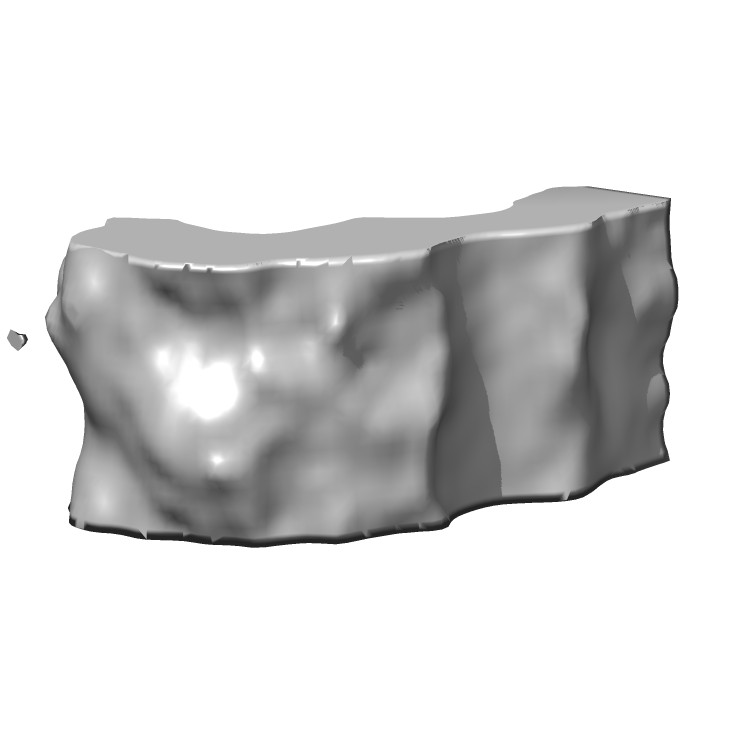 emd_5559_1.jpg emd_5559_1.jpg | 30 KB | ||
| Archive directory |  http://ftp.pdbj.org/pub/emdb/structures/EMD-5559ftp://ftp.pdbj.org/pub/emdb/structures/EMD-5559 http://ftp.pdbj.org/pub/emdb/structures/EMD-5559ftp://ftp.pdbj.org/pub/emdb/structures/EMD-5559 | HTTPS FTP |
-Validation report
| Summary document | emd_5559_validation.pdf.gz | 266.7 KB | Display | EMDB validaton report |
|---|---|---|---|---|
| Full document | emd_5559_full_validation.pdf.gz | 266.2 KB | Display | |
| Data in XML | emd_5559_validation.xml.gz | 4.2 KB | Display | |
| Arichive directory | https://ftp.pdbj.org/pub/emdb/validation_reports/EMD-5559ftp://ftp.pdbj.org/pub/emdb/validation_reports/EMD-5559 | HTTPS FTP |
-Related structure data
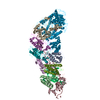
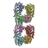
| Related structure data |  3j2sMC  5540C  3j2qC M: atomic model generated by this map C: citing same article ( |
|---|---|
| Similar structure data |


-Links
| EMDB pages | EMDB (EBI/PDBe) / EMDataResource |
|---|---|
| Related items in Molecule of the Month |


-Map
| File | Download / File: emd_5559.map.gz / Format: CCP4 / Size: 330.1 KB / Type: IMAGE STORED AS FLOATING POINT NUMBER (4 BYTES) | ||||||||||||||||||||||||||||||||||||||||||||||||||||||||||||||||||||
|---|---|---|---|---|---|---|---|---|---|---|---|---|---|---|---|---|---|---|---|---|---|---|---|---|---|---|---|---|---|---|---|---|---|---|---|---|---|---|---|---|---|---|---|---|---|---|---|---|---|---|---|---|---|---|---|---|---|---|---|---|---|---|---|---|---|---|---|---|---|
| Annotation | Cropped volume from helical reconstruction of membrane-bound Factor vIII light chain bound to single bilayer lipid nanotubes (EMD-5540) with a Gaussian filter of 3.0 applied | ||||||||||||||||||||||||||||||||||||||||||||||||||||||||||||||||||||
| Projections & slices | Image control
Images are generated by Spider. generated in cubic-lattice coordinate | ||||||||||||||||||||||||||||||||||||||||||||||||||||||||||||||||||||
| Voxel size | X=Y=Z: 2.9 Å | ||||||||||||||||||||||||||||||||||||||||||||||||||||||||||||||||||||
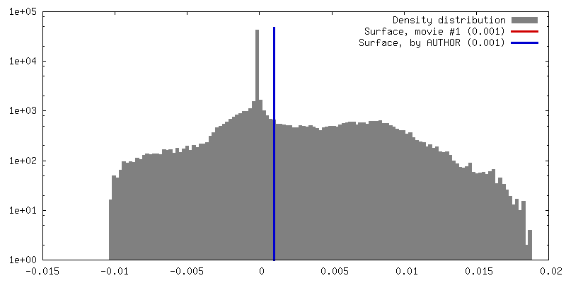
| Density |
| ||||||||||||||||||||||||||||||||||||||||||||||||||||||||||||||||||||
| Symmetry | Space group: 1 | ||||||||||||||||||||||||||||||||||||||||||||||||||||||||||||||||||||
| Details | EMDB XML:
CCP4 map header:
| ||||||||||||||||||||||||||||||||||||||||||||||||||||||||||||||||||||
 Z (Sec.)
 Y (Row.)
 X (Col.)


-Supplemental data
- Sample components
-Entire : Cropped volume corresponding to 2x1 factor VIII light chain molec...
| Entire | Name: Cropped volume corresponding to 2x1 factor VIII light chain molecules from EMD-5540 |
|---|---|
| Components |
|
-Supramolecule #1000: Cropped volume corresponding to 2x1 factor VIII light chain molec...
| Supramolecule | Name: Cropped volume corresponding to 2x1 factor VIII light chain molecules from EMD-5540 type: sample / ID: 1000 Oligomeric state: One molecule selected from 15 molecules organized around a 120-Angstrom lipid nanotube Number unique components: 1 |
|---|---|
| Molecular weight | Experimental: 80 KDa / Theoretical: 80 KDa / Method: SDS-PAGE |
-Macromolecule #1: blood coagulation Factor VIII light chain
| Macromolecule | Name: blood coagulation Factor VIII light chain / type: protein_or_peptide / ID: 1 / Name.synonym: Hemophilia factor light chain A Details: 15 molecules organized helically around a 120-Angstrom lipid nanotube Oligomeric state: 15 subunits helically organized onto lipid nanotube with a length of 114 Angstrom Recombinant expression: Yes |
|---|---|
| Source (natural) | Organism: Homo sapiens (human) / synonym: human / Location in cell: blood plasma |
| Molecular weight | Experimental: 80 KDa / Theoretical: 80 KDa |
| Recombinant expression | Organism:   Cricetulus griseus (Chinese hamster) / Recombinant cell: CHO Cricetulus griseus (Chinese hamster) / Recombinant cell: CHO |
| Sequence | UniProtKB: Coagulation factor VIII |
-Experimental details
-Structure determination
| Method | cryo EM |
|---|---|
 Processing Processing | helical reconstruction |
| Aggregation state | helical array |
-Sample preparation
| Concentration | 1 mg/mL |
|---|---|
| Buffer | pH: 7.4 / Details: 20 mM Tris-HCl, 150 mM NaCl, 5mM CaCl2 |
| Grid | Details: 300 mesh R2x2 Quantifoil grids |
| Vitrification | Cryogen name: ETHANE / Chamber humidity: 100 % / Chamber temperature: 95 K / Instrument: FEI VITROBOT MARK III / Method: Blot 3.5 seconds before plunging |
| Details | The protein was mixed in 1:1 w/w ratio with lipid nanotubes solution |
- Electron microscopy
| Microscope | JEOL 2010F |
|---|---|
| Temperature | Min: 93 K / Max: 103 K / Average: 99 K |
| Alignment procedure | Legacy - Astigmatism: corrected at 400,000 times magnification |
| Date | Apr 2, 2010 |
| Image recording | Category: CCD / Film or detector model: GATAN ULTRASCAN 4000 (4k x 4k) / Digitization - Sampling interval: 15 µm / Number real images: 69 / Average electron dose: 16 e/Å2 / Details: Each image was acquired for 1 second. |
| Electron beam | Acceleration voltage: 200 kV / Electron source:  FIELD EMISSION GUN FIELD EMISSION GUN |
| Electron optics | Calibrated magnification: 52000 / Illumination mode: FLOOD BEAM / Imaging mode: BRIGHT FIELD / Cs: 2.0 mm / Nominal defocus max: -4.4 µm / Nominal defocus min: -0.7 µm / Nominal magnification: 52000 |
| Sample stage | Specimen holder model: GATAN LIQUID NITROGEN |
-Image processing
| Details | IHRSR, SPIDER and EMAN2 |
|---|---|
| Final reconstruction | Applied symmetry - Helical parameters - Δz: 7.6 Å Applied symmetry - Helical parameters - Δ&Phi: 0.5 ° Algorithm: OTHER / Resolution.type: BY AUTHOR / Resolution: 15.0 Å / Resolution method: OTHER / Software - Name: IHRS, SPIDER, EMAN2 Details: The final 3D reconstructions was calculated from a set of 2043 helical segments cut off from the selected helical tubes at 256 x 256 pixels with 10% overlap. |
| CTF correction | Details: particle stacks for each micrograph were corrected for CTF (only phase correction) |
-Atomic model buiding 1
| Initial model | PDB ID: Chain - Chain ID: B |
|---|---|
| Software | Name: UCSF-Chimera, VMD |
| Details | The 3CDZ chain B coordinates were fitted flexibly within the 3D map with the 'fit to volume' option of the UCSF Chimera software. |
| Refinement | Space: REAL / Protocol: FLEXIBLE FIT / Target criteria: optimal fit |
| Output model | PDB-3j2s: |

