 ムービー
ムービー コントローラー


+ データを開く

- 基本情報
基本情報
| 登録情報 | データベース: EMDB / ID: EMD-4440 | ||||||||||||
|---|---|---|---|---|---|---|---|---|---|---|---|---|---|
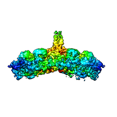
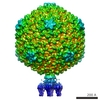
| タイトル | Structure of head fiber and inner core protein gp22 of native bacteriophage P68 | ||||||||||||
 マップデータ マップデータ | Map of head fiber and gp22 bound to hexon adjacent to portal, masked from five-fold cryoEM reconstruction of native bacteriophage P68 capsid | ||||||||||||
 試料 試料 |
| ||||||||||||
 キーワード キーワード | structural protein / bacteriophage / head fiber / inner core protein | ||||||||||||
| 機能・相同性 | Uncharacterized protein / Major head protein 機能・相同性情報 機能・相同性情報 | ||||||||||||
| 生物種 |  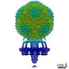 Staphylococcus phage P68 (ファージ) Staphylococcus phage P68 (ファージ) | ||||||||||||
| 手法 | 単粒子再構成法 / クライオ電子顕微鏡法 / 解像度: 3.8 Å | ||||||||||||
 データ登録者 データ登録者 | Hrebik D / Skubnik K / Fuzik T / Plevka P | ||||||||||||
| 資金援助 |  チェコ, 3件 チェコ, 3件
| ||||||||||||
 引用 引用 | ジャーナル: Sci Adv / 年: 2019 タイトル: Structure and genome ejection mechanism of phage P68. 著者: Dominik Hrebík / Dana Štveráková / Karel Škubník / Tibor Füzik / Roman Pantůček / Pavel Plevka / 要旨: Phages infecting can be used as therapeutics against antibiotic-resistant bacterial infections. However, there is limited information about the mechanism of genome delivery of phages that infect ...Phages infecting can be used as therapeutics against antibiotic-resistant bacterial infections. However, there is limited information about the mechanism of genome delivery of phages that infect Gram-positive bacteria. Here, we present the structures of native phage P68, genome ejection intermediate, and empty particle. The P68 head contains 72 subunits of inner core protein, 15 of which bind to and alter the structure of adjacent major capsid proteins and thus specify attachment sites for head fibers. Unlike in the previously studied phages, the head fibers of P68 enable its virion to position itself at the cell surface for genome delivery. The unique interaction of one end of P68 DNA with one of the 12 portal protein subunits is disrupted before the genome ejection. The inner core proteins are released together with the DNA and enable the translocation of phage genome across the bacterial membrane into the cytoplasm. | ||||||||||||
| 履歴 |
|
- 構造の表示
構造の表示
| ムービー |
ムービービューア |
|---|---|
| 構造ビューア | EMマップ: SurfViewMolmilJmol/JSmol |
| 添付画像 |
- ダウンロードとリンク
-EMDBアーカイブ
| マップデータ | emd_4440.map.gz | 2 MB | EMDBマップデータ形式 | |
|---|---|---|---|---|
| ヘッダ (付随情報) | emd-4440-v30.xmlemd-4440.xml | 19.5 KB 19.5 KB | 表示 表示 | EMDBヘッダ |
| 画像 |  emd_4440.png emd_4440.png | 81.1 KB | ||
| Filedesc metadata | emd-4440.cif.gz | 6.9 KB | ||
| アーカイブディレクトリ |  http://ftp.pdbj.org/pub/emdb/structures/EMD-4440ftp://ftp.pdbj.org/pub/emdb/structures/EMD-4440 http://ftp.pdbj.org/pub/emdb/structures/EMD-4440ftp://ftp.pdbj.org/pub/emdb/structures/EMD-4440 | HTTPS FTP |
-関連構造データ
| 関連構造データ |  6iawMC  4435C 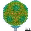 4436C  4437C  4438C 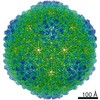 4442C  4449C  4450C 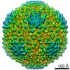 4451C  4453C  4454C  4455C 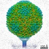 4456C  4457C  4458C 4459C  6iabC 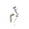 6iacC  6iatC  6ib1C  6q3gC M: このマップから作成された原子モデル C: 同じ文献を引用 ( |
|---|---|
| 類似構造データ |

-リンク
| EMDBのページ | EMDB (EBI/PDBe) / EMDataResource |
|---|
-マップ
| ファイル | ダウンロード / ファイル: emd_4440.map.gz / 形式: CCP4 / 大きさ: 30.5 MB / タイプ: IMAGE STORED AS FLOATING POINT NUMBER (4 BYTES) | ||||||||||||||||||||||||||||||||||||||||||||||||||||||||||||
|---|---|---|---|---|---|---|---|---|---|---|---|---|---|---|---|---|---|---|---|---|---|---|---|---|---|---|---|---|---|---|---|---|---|---|---|---|---|---|---|---|---|---|---|---|---|---|---|---|---|---|---|---|---|---|---|---|---|---|---|---|---|
| 注釈 | Map of head fiber and gp22 bound to hexon adjacent to portal, masked from five-fold cryoEM reconstruction of native bacteriophage P68 capsid | ||||||||||||||||||||||||||||||||||||||||||||||||||||||||||||
| 投影像・断面図 | 画像のコントロール
画像は Spider により作成 | ||||||||||||||||||||||||||||||||||||||||||||||||||||||||||||
| ボクセルのサイズ | X=Y=Z: 1.063 Å | ||||||||||||||||||||||||||||||||||||||||||||||||||||||||||||
| 密度 |
| ||||||||||||||||||||||||||||||||||||||||||||||||||||||||||||
| 対称性 | 空間群: 1 | ||||||||||||||||||||||||||||||||||||||||||||||||||||||||||||
| 詳細 | EMDB XML:
CCP4マップ ヘッダ情報:
| ||||||||||||||||||||||||||||||||||||||||||||||||||||||||||||
 Z (Sec.)
 Y (Row.)
 X (Col.)


-添付データ
- 試料の構成要素
-全体 : Staphylococcus phage P68
| 全体 | 名称: Staphylococcus phage P68 (ファージ) |
|---|---|
| 要素 |
|
-超分子 #1: Staphylococcus phage P68
| 超分子 | 名称: Staphylococcus phage P68 / タイプ: virus / ID: 1 / 親要素: 0 / 含まれる分子: all / NCBI-ID: 204090 / 生物種: Staphylococcus phage P68 / ウイルスタイプ: VIRION / ウイルス・単離状態: STRAIN / ウイルス・エンベロープ: No / ウイルス・中空状態: No |
|---|---|
| 宿主 | 生物種:   Staphylococcus aureus (黄色ブドウ球菌) / 株: dTarM 4220 Staphylococcus aureus (黄色ブドウ球菌) / 株: dTarM 4220 |
| 分子量 | 理論値: 19.7 MDa |
| ウイルス殻 | Shell ID: 1 / 名称: Capsid / 直径: 480.0 Å / T番号(三角分割数): 4 |
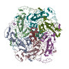
-分子 #1: Major head protein
| 分子 | 名称: Major head protein / タイプ: protein_or_peptide / ID: 1 / コピー数: 6 / 光学異性体: LEVO |
|---|---|
| 由来(天然) | 生物種: Staphylococcus phage P68 (ファージ) |
| 分子量 | 理論値: 46.954941 KDa |
| 配列 | 文字列: MAQQSTKNET ALLVAKSAKS ALQDFNHDYS KSWTFGDKWD NSNTMFETFV NKYLFPKINE TLLIDIALGN RFNWLAKEQD FIGQYSEEY VIMDTVPINM DLSKNEELML KRNYPRMATK LYGNGIVKKQ KFTLNNNDTR FNFQTLADAT NYALGVYKKK I SDINVLEE ...文字列: MAQQSTKNET ALLVAKSAKS ALQDFNHDYS KSWTFGDKWD NSNTMFETFV NKYLFPKINE TLLIDIALGN RFNWLAKEQD FIGQYSEEY VIMDTVPINM DLSKNEELML KRNYPRMATK LYGNGIVKKQ KFTLNNNDTR FNFQTLADAT NYALGVYKKK I SDINVLEE KEMRAMLVDY SLNQLSETNV RKATSKEDLA SKVFEAILNL QNNSAKYNEV HRASGGAIGQ YTTVSKLKDI VI LTTDSLK SYLLDTKIAN TFQIAGIDFT DHVISFDDLG GVFKVTKEFK LQNQDSIDFL RAYGDYQSQL GDTIPVGAVF TYD VSKLKE FTGNVEEIKP KSDLYAFILD INSIKYKRYT KGMLKPPFHN PEFDEVTHWI HYYSFKAISP FFNKILITDQ DVNP KPEEE LQE UniProtKB: Major head protein |
-分子 #2: Arstotzka protein
| 分子 | 名称: Arstotzka protein / タイプ: protein_or_peptide / ID: 2 / コピー数: 6 / 光学異性体: LEVO |
|---|---|
| 由来(天然) | 生物種: Staphylococcus phage P68 (ファージ) |
| 分子量 | 理論値: 6.922464 KDa |
| 配列 | 文字列: MYEGNNMRSM MGTSYEDSRL NKRTELNENM SIDTNKSEDS YGVQIHSLSK QSFTGDVEEE UniProtKB: Uncharacterized protein |
-分子 #3: Head fiber
| 分子 | 名称: Head fiber / タイプ: protein_or_peptide / ID: 3 詳細: Poly alanine chain fitted to the density of head-fiber コピー数: 3 / 光学異性体: LEVO |
|---|---|
| 由来(天然) | 生物種: Staphylococcus phage P68 (ファージ) |
| 分子量 | 理論値: 5.720042 KDa |
| 配列 | 文字列: (UNK)(UNK)(UNK)(UNK)(UNK)(UNK)(UNK)(UNK)(UNK)(UNK) (UNK)(UNK)(UNK)(UNK)(UNK)(UNK) (UNK)(UNK)(UNK) (UNK)(UNK)(UNK)(UNK)(UNK)(UNK)(UNK)(UNK)(UNK)(UNK) (UNK)(UNK)(UNK) (UNK)(UNK)(UNK)(UNK) ...文字列: (UNK)(UNK)(UNK)(UNK)(UNK)(UNK)(UNK)(UNK)(UNK)(UNK) (UNK)(UNK)(UNK)(UNK)(UNK)(UNK) (UNK)(UNK)(UNK) (UNK)(UNK)(UNK)(UNK)(UNK)(UNK)(UNK)(UNK)(UNK)(UNK) (UNK)(UNK)(UNK) (UNK)(UNK)(UNK)(UNK)(UNK)(UNK) (UNK)(UNK)(UNK)(UNK)(UNK)(UNK)(UNK)(UNK)(UNK)(UNK) (UNK)(UNK)(UNK)(UNK)(UNK)(UNK)(UNK)(UNK)(UNK) (UNK)(UNK)(UNK)(UNK)(UNK)(UNK)(UNK) (UNK)(UNK) (UNK) |
-分子 #4: inner core protein
| 分子 | 名称: inner core protein / タイプ: protein_or_peptide / ID: 4 詳細: Poly alanine chain fitted to the density of the inner core protein bound to hexon adjacent to portal コピー数: 3 / 光学異性体: LEVO |
|---|---|
| 由来(天然) | 生物種: Staphylococcus phage P68 (ファージ) |
| 分子量 | 理論値: 3.507314 KDa |
| 配列 | 文字列: (UNK)(UNK)(UNK)(UNK)(UNK)(UNK)(UNK)(UNK)(UNK)(UNK) (UNK)(UNK)(UNK)(UNK)(UNK)(UNK) (UNK)(UNK)(UNK) (UNK)(UNK)(UNK)(UNK)(UNK)(UNK)(UNK)(UNK)(UNK)(UNK) (UNK)(UNK)(UNK) (UNK)(UNK)(UNK)(UNK) ...文字列: (UNK)(UNK)(UNK)(UNK)(UNK)(UNK)(UNK)(UNK)(UNK)(UNK) (UNK)(UNK)(UNK)(UNK)(UNK)(UNK) (UNK)(UNK)(UNK) (UNK)(UNK)(UNK)(UNK)(UNK)(UNK)(UNK)(UNK)(UNK)(UNK) (UNK)(UNK)(UNK) (UNK)(UNK)(UNK)(UNK)(UNK)(UNK) (UNK)(UNK)(UNK) |
-実験情報
-構造解析
| 手法 | クライオ電子顕微鏡法 |
|---|---|
 解析 解析 | 単粒子再構成法 |
| 試料の集合状態 | particle |
-試料調製
| 濃度 | 2 mg/mL | ||||||||||||
|---|---|---|---|---|---|---|---|---|---|---|---|---|---|
| 緩衝液 | pH: 8 構成要素:
| ||||||||||||
| グリッド | モデル: Quantifoil R2/1 / 材質: COPPER / メッシュ: 200 / 支持フィルム - 材質: CARBON / 支持フィルム - トポロジー: HOLEY / 前処理 - タイプ: GLOW DISCHARGE / 前処理 - 時間: 30 sec. / 前処理 - 雰囲気: NITROGEN | ||||||||||||
| 凍結 | 凍結剤: ETHANE / チャンバー内湿度: 100 % / チャンバー内温度: 293 K / 装置: FEI VITROBOT MARK IV / 詳細: blot time 2s; blot force -2; 3.6 ul of sample. |
- 電子顕微鏡法
| 顕微鏡 | FEI TITAN KRIOS |
|---|---|
| 撮影 | フィルム・検出器のモデル: FEI FALCON II (4k x 4k) 検出モード: INTEGRATING / デジタル化 - サイズ - 横: 4096 pixel / デジタル化 - サイズ - 縦: 4096 pixel / デジタル化 - 画像ごとのフレーム数: 1-7 / 撮影したグリッド数: 2 / 実像数: 2891 / 平均露光時間: 1.0 sec. / 平均電子線量: 21.0 e/Å2 |
| 電子線 | 加速電圧: 300 kV / 電子線源:  FIELD EMISSION GUN FIELD EMISSION GUN |
| 電子光学系 | C2レンズ絞り径: 70.0 µm / 照射モード: FLOOD BEAM / 撮影モード: BRIGHT FIELD / Cs: 2.7 mm / 最大 デフォーカス(公称値): 0.003 µm / 最小 デフォーカス(公称値): 0.001 µm / 倍率(公称値): 75000 |
| 試料ステージ | 試料ホルダーモデル: FEI TITAN KRIOS AUTOGRID HOLDER ホルダー冷却材: NITROGEN |
| 実験機器 |  モデル: Titan Krios / 画像提供: FEI Company |
+画像解析
-原子モデル構築 1
| 初期モデル | PDB ID: Chain - Source name: PDB / Chain - Initial model type: experimental model |
|---|---|
| 詳細 | The refinement was conducted on rigid body fitted major capsid proteins and Arstotzka proteins obtained from related structure of 6IAT; EMD-4438. The chains were fitted into hexon adjacent to portal of P68 capsid obtained from 5-fold symmetrized reconstruction. Subsequently, poly-alanine chains were manually built into densities corresponding to N-terminal part of head-fiber (chain IDs H,K,L) and C-terminal part of inner core protein (chain IDs X,Y,Z). The map was masked according to the related pdb in chimera, normalized and put into several rounds of a real space refinement in Phenix. |
| 精密化 | 空間: REAL / プロトコル: AB INITIO MODEL |
| 得られたモデル | PDB-6iaw: |
