 ムービー
ムービー コントローラー


+ データを開く

- 基本情報
基本情報
| 登録情報 | データベース: PDB / ID: 6iaw | |||||||||||||||
|---|---|---|---|---|---|---|---|---|---|---|---|---|---|---|---|---|
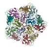
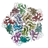
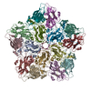
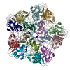
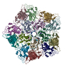
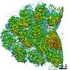
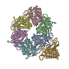
| タイトル | Structure of head fiber and inner core protein gp22 of native bacteriophage P68 | |||||||||||||||
 要素 要素 |
| |||||||||||||||
 キーワード キーワード | STRUCTURAL PROTEIN / bacteriophage / head fiber / inner core protein | |||||||||||||||
| 機能・相同性 | Uncharacterized protein / Major head protein 機能・相同性情報 機能・相同性情報 | |||||||||||||||
| 生物種 |  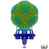 Staphylococcus phage P68 (ファージ) Staphylococcus phage P68 (ファージ) | |||||||||||||||
| 手法 | 電子顕微鏡法 / 単粒子再構成法 / クライオ電子顕微鏡法 / 解像度: 3.8 Å | |||||||||||||||
 データ登録者 データ登録者 | Hrebik, D. / Skubnik, K. / Fuzik, T. / Plevka, P. | |||||||||||||||
| 資金援助 |  チェコ, 4件 チェコ, 4件
| |||||||||||||||
 引用 引用 | ジャーナル: Sci Adv / 年: 2019 タイトル: Structure and genome ejection mechanism of phage P68. 著者: Dominik Hrebík / Dana Štveráková / Karel Škubník / Tibor Füzik / Roman Pantůček / Pavel Plevka / 要旨: Phages infecting can be used as therapeutics against antibiotic-resistant bacterial infections. However, there is limited information about the mechanism of genome delivery of phages that infect ...Phages infecting can be used as therapeutics against antibiotic-resistant bacterial infections. However, there is limited information about the mechanism of genome delivery of phages that infect Gram-positive bacteria. Here, we present the structures of native phage P68, genome ejection intermediate, and empty particle. The P68 head contains 72 subunits of inner core protein, 15 of which bind to and alter the structure of adjacent major capsid proteins and thus specify attachment sites for head fibers. Unlike in the previously studied phages, the head fibers of P68 enable its virion to position itself at the cell surface for genome delivery. The unique interaction of one end of P68 DNA with one of the 12 portal protein subunits is disrupted before the genome ejection. The inner core proteins are released together with the DNA and enable the translocation of phage genome across the bacterial membrane into the cytoplasm. | |||||||||||||||
| 履歴 |
|
- 構造の表示
構造の表示
| ムービー |
ムービービューア |
|---|---|
| 構造ビューア | 分子: MolmilJmol/JSmol |
- ダウンロードとリンク
-ダウンロード
| PDBx/mmCIF形式 | 6iaw.cif.gz | 475 KB | 表示 | PDBx/mmCIF形式 |
|---|---|---|---|---|
| PDB形式 | pdb6iaw.ent.gz | 393.2 KB | 表示 | PDB形式 |
| PDBx/mmJSON形式 | 6iaw.json.gz | ツリー表示 | PDBx/mmJSON形式 | |
| その他 |  その他のダウンロード その他のダウンロード |
-検証レポート
| アーカイブディレクトリ | https://data.pdbj.org/pub/pdb/validation_reports/ia/6iawftp://data.pdbj.org/pub/pdb/validation_reports/ia/6iaw | HTTPS FTP |
|---|
-関連構造データ
| 関連構造データ |  4440MC  4435C 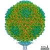 4436C  4437C  4438C 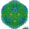 4442C  4449C  4450C 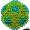 4451C  4453C  4454C  4455C 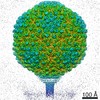 4456C  4457C  4458C 4459C  6iabC 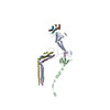 6iacC  6iatC  6ib1C  6q3gC M: このデータのモデリングに利用したマップデータ C: 同じ文献を引用 ( |
|---|---|
| 類似構造データ |
-リンク
 PDBj
PDBj- 集合体
集合体
| 登録構造単位 | 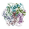
|
|---|---|
| 1 |
|
-要素
| #1: タンパク質 | 分子量: 46954.941 Da / 分子数: 6 / 由来タイプ: 天然 / 由来: (天然) Staphylococcus phage P68 (ファージ) / 参照: UniProt: Q859I3#2: タンパク質 | 分子量: 6922.464 Da / 分子数: 6 / 由来タイプ: 天然 / 由来: (天然) Staphylococcus phage P68 (ファージ) / 参照: UniProt: Q859I2#3: タンパク質 | 分子量: 5720.042 Da / 分子数: 3 / 由来タイプ: 天然 詳細: Poly alanine chain fitted to the density of head-fiber 由来: (天然) Staphylococcus phage P68 (ファージ)#4: タンパク質・ペプチド | 分子量: 3507.314 Da / 分子数: 3 / 由来タイプ: 天然 詳細: Poly alanine chain fitted to the density of the inner core protein bound to hexon adjacent to portal 由来: (天然) Staphylococcus phage P68 (ファージ) |
|---|
-実験情報
-実験
| 実験 | 手法: 電子顕微鏡法 |
|---|---|
| EM実験 | 試料の集合状態: PARTICLE / 3次元再構成法: 単粒子再構成法 |
- 試料調製
試料調製
| 構成要素 | 名称: Staphylococcus phage P68 / タイプ: VIRUS / Entity ID: all / 由来: NATURAL | ||||||||||||||||||||
|---|---|---|---|---|---|---|---|---|---|---|---|---|---|---|---|---|---|---|---|---|---|
| 分子量 | 値: 19.7 MDa / 実験値: NO | ||||||||||||||||||||
| 由来(天然) | 生物種: Staphylococcus phage P68 (ファージ) | ||||||||||||||||||||
| ウイルスについての詳細 | 中空か: NO / エンベロープを持つか: NO / 単離: STRAIN / タイプ: VIRION | ||||||||||||||||||||
| 天然宿主 | 生物種: Staphylococcus aureus / 株: dTarM 4220 | ||||||||||||||||||||
| ウイルス殻 | 名称: Capsid / 直径: 480 nm / 三角数 (T数): 4 | ||||||||||||||||||||
| 緩衝液 | pH: 8 | ||||||||||||||||||||
| 緩衝液成分 |
| ||||||||||||||||||||
| 試料 | 濃度: 2 mg/ml / 包埋: NO / シャドウイング: NO / 染色: NO / 凍結: YES | ||||||||||||||||||||
| 試料支持 | グリッドの材料: COPPER / グリッドのサイズ: 200 divisions/in. / グリッドのタイプ: Quantifoil R2/1 | ||||||||||||||||||||
| 急速凍結 | 装置: FEI VITROBOT MARK IV / 凍結剤: ETHANE / 湿度: 100 % / 凍結前の試料温度: 293 K / 詳細: blot time 2s; blot force -2; 3.6 ul of sample |
- 電子顕微鏡撮影
| 実験機器 |  モデル: Titan Krios / 画像提供: FEI Company |
|---|---|
| 顕微鏡 | モデル: FEI TITAN KRIOS |
| 電子銃 | 電子線源:  FIELD EMISSION GUN / 加速電圧: 300 kV / 照射モード: FLOOD BEAM FIELD EMISSION GUN / 加速電圧: 300 kV / 照射モード: FLOOD BEAM |
| 電子レンズ | モード: BRIGHT FIELD / 倍率(公称値): 75000 X / 最大 デフォーカス(公称値): 3 nm / 最小 デフォーカス(公称値): 1 nm / Cs: 2.7 mm / C2レンズ絞り径: 70 µm / アライメント法: COMA FREE |
| 試料ホルダ | 凍結剤: NITROGEN 試料ホルダーモデル: FEI TITAN KRIOS AUTOGRID HOLDER |
| 撮影 | 平均露光時間: 1 sec. / 電子線照射量: 21 e/Å2 / 検出モード: INTEGRATING フィルム・検出器のモデル: FEI FALCON II (4k x 4k) 撮影したグリッド数: 2 / 実像数: 2891 |
| 画像スキャン | 横: 4096 / 縦: 4096 / 動画フレーム数/画像: 7 / 利用したフレーム数/画像: 1-7 |
- 解析
解析
| EMソフトウェア |
| ||||||||||||||||||||||||||||||||||||||||
|---|---|---|---|---|---|---|---|---|---|---|---|---|---|---|---|---|---|---|---|---|---|---|---|---|---|---|---|---|---|---|---|---|---|---|---|---|---|---|---|---|---|
| CTF補正 | タイプ: PHASE FLIPPING AND AMPLITUDE CORRECTION | ||||||||||||||||||||||||||||||||||||||||
| 粒子像の選択 | 選択した粒子像数: 37218 | ||||||||||||||||||||||||||||||||||||||||
| 対称性 | 点対称性: C5 (5回回転対称) | ||||||||||||||||||||||||||||||||||||||||
| 3次元再構成 | 解像度: 3.8 Å / 解像度の算出法: FSC 0.143 CUT-OFF / 粒子像の数: 28826 / アルゴリズム: BACK PROJECTION / クラス平均像の数: 1 / 対称性のタイプ: POINT | ||||||||||||||||||||||||||||||||||||||||
| 原子モデル構築 | プロトコル: AB INITIO MODEL / 空間: REAL 詳細: The refinement was conducted on rigid body fitted major capsid proteins and Arstotzka proteins obtained from related structure of 6IAT; EMD-4438. The chains were fitted into hexon adjacent to ...詳細: The refinement was conducted on rigid body fitted major capsid proteins and Arstotzka proteins obtained from related structure of 6IAT; EMD-4438. The chains were fitted into hexon adjacent to portal of P68 capsid obtained from 5-fold symmetrized reconstruction. Subsequently, poly-alanine chains were manually built into densities corresponding to N-terminal part of head-fiber (chain IDs H,K,L) and C-terminal part of inner core protein (chain IDs X,Y,Z). The map was masked according to the related pdb in chimera, normalized and put into several rounds of a real space refinement in Phenix. | ||||||||||||||||||||||||||||||||||||||||
| 原子モデル構築 | PDB-ID: 6IAT Accession code: 6IAT / Source name: PDB / タイプ: experimental model |
