 Movie
Movie Controller


+ Open data

- Basic information
| Entry | Database: PDB / ID: 6iab | |||||||||||||||
|---|---|---|---|---|---|---|---|---|---|---|---|---|---|---|---|---|
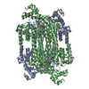
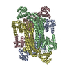
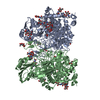
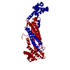
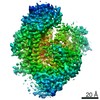
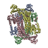
| Title | Tail fiber of Staphylococcus aureus phage P68 | |||||||||||||||
 Components Components | Tail Fiber of phage P68 | |||||||||||||||
 Keywords Keywords | SUGAR BINDING PROTEIN / structural protein / receptor binding protein / beta-propeller | |||||||||||||||
| Function / homology | : / Phage 5-bladed beta propeller receptor binding platform domain / Minor structural protein / Minor structural protein Function and homology information Function and homology information | |||||||||||||||
| Biological species |  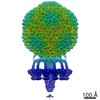 Staphylococcus aureus phage P68 (virus) Staphylococcus aureus phage P68 (virus) | |||||||||||||||
| Method |  X-RAY DIFFRACTION / SYNCHROTRON / MOLECULAR REPLACEMENT / Resolution: 2 Å X-RAY DIFFRACTION / SYNCHROTRON / MOLECULAR REPLACEMENT / Resolution: 2 Å | |||||||||||||||
 Authors Authors | Hrebik, D. / Karel, S. / Plevka, P. | |||||||||||||||
| Funding support |  Czech Republic, 4items Czech Republic, 4items
| |||||||||||||||
 Citation Citation | Journal: Sci Adv / Year: 2019 Title: Structure and genome ejection mechanism of phage P68. Authors: Dominik Hrebík / Dana Štveráková / Karel Škubník / Tibor Füzik / Roman Pantůček / Pavel Plevka / Abstract: Phages infecting can be used as therapeutics against antibiotic-resistant bacterial infections. However, there is limited information about the mechanism of genome delivery of phages that infect ...Phages infecting can be used as therapeutics against antibiotic-resistant bacterial infections. However, there is limited information about the mechanism of genome delivery of phages that infect Gram-positive bacteria. Here, we present the structures of native phage P68, genome ejection intermediate, and empty particle. The P68 head contains 72 subunits of inner core protein, 15 of which bind to and alter the structure of adjacent major capsid proteins and thus specify attachment sites for head fibers. Unlike in the previously studied phages, the head fibers of P68 enable its virion to position itself at the cell surface for genome delivery. The unique interaction of one end of P68 DNA with one of the 12 portal protein subunits is disrupted before the genome ejection. The inner core proteins are released together with the DNA and enable the translocation of phage genome across the bacterial membrane into the cytoplasm. | |||||||||||||||
| History |
|
- Structure visualization
| Structure viewer | Molecule: MolmilJmol/JSmol |
|---|
- Downloads & links
-Download
| PDBx/mmCIF format | 6iab.cif.gz | 128.5 KB | Display | PDBx/mmCIF format |
|---|---|---|---|---|
| PDB format | pdb6iab.ent.gz | 95.3 KB | Display | PDB format |
| PDBx/mmJSON format | 6iab.json.gz | Tree view | PDBx/mmJSON format | |
| Others |  Other downloads Other downloads |
-Validation report
| Arichive directory | https://data.pdbj.org/pub/pdb/validation_reports/ia/6iabftp://data.pdbj.org/pub/pdb/validation_reports/ia/6iab | HTTPS FTP |
|---|
-Related structure data
| Related structure data |  4435C 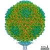 4436C  4437C  4438C  4440C 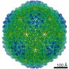 4442C  4449C  4450C 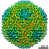 4451C  4453C  4454C  4455C 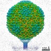 4456C  4457C  4458C 4459C 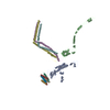 6iacC 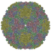 6iatC 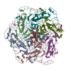 6iawC 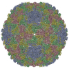 6ib1C 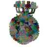 6q3gC  5efvS S: Starting model for refinement C: citing same article ( |
|---|---|
| Similar structure data |


-Links
 PDBj
PDBj- Assembly
| Deposited unit | 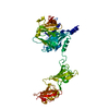
| |||||||||||||||||||||
|---|---|---|---|---|---|---|---|---|---|---|---|---|---|---|---|---|---|---|---|---|---|---|
| 1 | 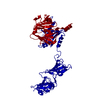
| |||||||||||||||||||||
| Unit cell |
| |||||||||||||||||||||
| Components on special symmetry positions |
|
-Components
| #1: Protein | Mass: 75662.641 Da / Num. of mol.: 1 Source method: isolated from a genetically manipulated source Source: (gene. exp.) Staphylococcus aureus phage P68 (virus)Plasmid: pMUH17 / Production host:  |
|---|---|
| #2: Water | ChemComp-HOH /  Mass: 18.015 Da / Num. of mol.: 436 / Source method: isolated from a natural source / Formula: H2O Mass: 18.015 Da / Num. of mol.: 436 / Source method: isolated from a natural source / Formula: H2O |
-Experimental details
-Experiment
| Experiment | Method: X-RAY DIFFRACTION / Number of used crystals: 1 |
|---|
- Sample preparation
| Crystal | Density Matthews: 1.62 Å3/Da / Density % sol: 24.18 % |
|---|---|
| Crystal grow | Temperature: 293.16 K / Method: vapor diffusion, hanging drop Details: 200 mM magnesium chloride, 100 mM HEPES, 20% (w/v) polyethylene glycol 6000 PH range: 7.0 - 7.5 |
-Data collection
| Diffraction | Mean temperature: 80 K / Serial crystal experiment: N |
|---|---|
| Diffraction source | Source: SYNCHROTRON / Site: SOLEIL  / Beamline: PROXIMA 1 / Wavelength: 0.97857 Å / Beamline: PROXIMA 1 / Wavelength: 0.97857 Å |
| Detector | Type: DECTRIS PILATUS 6M / Detector: PIXEL / Date: May 19, 2017 |
| Radiation | Monochromator: channel cut cryogenically cooled monochromator crystal Protocol: SINGLE WAVELENGTH / Monochromatic (M) / Laue (L): M / Scattering type: x-ray |
| Radiation wavelength | Wavelength: 0.97857 Å / Relative weight: 1 |
| Reflection | Resolution: 2→19.968 Å / Num. obs: 32065 / % possible obs: 99.99 % / Redundancy: 5.1 % / Net I/σ(I): 13.5 |
| Reflection shell | Resolution: 2.0002→2.0502 Å / Num. unique obs: 2146 / % possible all: 100 |
- Processing
| Software |
| |||||||||||||||||||||||||||||||||||||||||||||||||||||||||||||||||||||||||||||||||||||||||||||||||||||||||
|---|---|---|---|---|---|---|---|---|---|---|---|---|---|---|---|---|---|---|---|---|---|---|---|---|---|---|---|---|---|---|---|---|---|---|---|---|---|---|---|---|---|---|---|---|---|---|---|---|---|---|---|---|---|---|---|---|---|---|---|---|---|---|---|---|---|---|---|---|---|---|---|---|---|---|---|---|---|---|---|---|---|---|---|---|---|---|---|---|---|---|---|---|---|---|---|---|---|---|---|---|---|---|---|---|---|---|
| Refinement | Method to determine structure: MOLECULAR REPLACEMENT Starting model: 5EFV Resolution: 2→19.968 Å / SU ML: 0.17 / Cross valid method: FREE R-VALUE / σ(F): 1.59 / Phase error: 18.9
| |||||||||||||||||||||||||||||||||||||||||||||||||||||||||||||||||||||||||||||||||||||||||||||||||||||||||
| Solvent computation | Shrinkage radii: 0.9 Å / VDW probe radii: 1.11 Å | |||||||||||||||||||||||||||||||||||||||||||||||||||||||||||||||||||||||||||||||||||||||||||||||||||||||||
| Refinement step | Cycle: LAST / Resolution: 2→19.968 Å
| |||||||||||||||||||||||||||||||||||||||||||||||||||||||||||||||||||||||||||||||||||||||||||||||||||||||||
| Refine LS restraints |
| |||||||||||||||||||||||||||||||||||||||||||||||||||||||||||||||||||||||||||||||||||||||||||||||||||||||||
| LS refinement shell |
|
