[English] 日本語
 Yorodumi
- PDB-3j3x: Independent reconstruction of Mm-cpn cryo-EM density map from hal... -
+ Open data
- Basic information
| Entry | Database: PDB / ID: 3j3x | ||||||
|---|---|---|---|---|---|---|---|
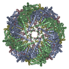
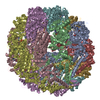
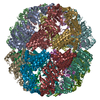
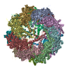
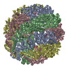
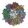
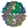
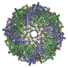
| Title | Independent reconstruction of Mm-cpn cryo-EM density map from half dataset in the closed state (training map) | ||||||
 Components Components | Chaperonin | ||||||
 Keywords Keywords | CHAPERONE / modeling / independent reconstruction / cryo-EM model validation | ||||||
| Function / homology |  Function and homology information Function and homology informationATP-dependent protein folding chaperone / unfolded protein binding / ATP hydrolysis activity / protein-containing complex / ATP binding / metal ion binding / identical protein binding / cytoplasm Similarity search - Function | ||||||
| Biological species |  Methanococcus maripaludis (archaea) Methanococcus maripaludis (archaea) | ||||||
| Method | ELECTRON MICROSCOPY / single particle reconstruction / cryo EM / Resolution: 4.3 Å | ||||||
 Authors Authors | DiMaio, F. / Zhang, J. / Chiu, W. / Baker, D. | ||||||
 Citation Citation |  Journal: Protein Sci / Year: 2013 Journal: Protein Sci / Year: 2013Title: Cryo-EM model validation using independent map reconstructions. Authors: Frank DiMaio / Junjie Zhang / Wah Chiu / David Baker /  Abstract: An increasing number of cryo-electron microscopy (cryo-EM) density maps are being generated with suitable resolution to trace the protein backbone and guide sidechain placement. Generating and ...An increasing number of cryo-electron microscopy (cryo-EM) density maps are being generated with suitable resolution to trace the protein backbone and guide sidechain placement. Generating and evaluating atomic models based on such maps would be greatly facilitated by independent validation metrics for assessing the fit of the models to the data. We describe such a metric based on the fit of atomic models with independent test maps from single particle reconstructions not used in model refinement. The metric provides a means to determine the proper balance between the fit to the density and model energy and stereochemistry during refinement, and is likely to be useful in determining values of model building and refinement metaparameters quite generally. | ||||||
| History |
|
- Structure visualization
| Movie |
 Movie viewer Movie viewer |
|---|---|
| Structure viewer | Molecule:  Molmil Molmil Jmol/JSmol Jmol/JSmol |
- Downloads & links
- Download
| PDBx/mmCIF format |  3j3x.cif.gz 3j3x.cif.gz | 1.2 MB | Display |  PDBx/mmCIF format PDBx/mmCIF format |
|---|---|---|---|---|
| PDB format |  pdb3j3x.ent.gz pdb3j3x.ent.gz | 1 MB | Display |  PDB format PDB format |
| PDBx/mmJSON format |  3j3x.json.gz 3j3x.json.gz | Tree view |  PDBx/mmJSON format PDBx/mmJSON format | |
| Others |  Other downloads Other downloads |
-Validation report
| Arichive directory |  https://data.pdbj.org/pub/pdb/validation_reports/j3/3j3x https://data.pdbj.org/pub/pdb/validation_reports/j3/3j3x ftp://data.pdbj.org/pub/pdb/validation_reports/j3/3j3x ftp://data.pdbj.org/pub/pdb/validation_reports/j3/3j3x | HTTPS FTP |
|---|
-Related structure data
| Related structure data |  5645MC  5646C M: map data used to model this data C: citing same article ( |
|---|---|
| Similar structure data |
- Links
Links
- Assembly
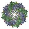
| Deposited unit | 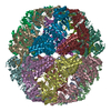
|
|---|---|
| 1 |
|
- Components
| #1: Protein | Mass: 57211.828 Da / Num. of mol.: 16 Source method: isolated from a genetically manipulated source Source: (gene. exp.)  Methanococcus maripaludis (archaea) / Production host: Methanococcus maripaludis (archaea) / Production host:  |
|---|
-Experimental details
-Experiment
| Experiment | Method: ELECTRON MICROSCOPY |
|---|---|
| EM experiment | Aggregation state: PARTICLE / 3D reconstruction method: single particle reconstruction |
- Sample preparation
| Component | Name: Mm-cpn with 1mM ATP/AlFx / Type: COMPLEX / Details: 16-mer |
|---|---|
| Molecular weight | Value: 0.96 MDa / Experimental value: NO |
| Specimen | Embedding applied: NO / Shadowing applied: NO / Staining applied: NO / Vitrification applied: YES |
| Vitrification | Instrument: FEI VITROBOT MARK III / Cryogen name: ETHANE / Humidity: 100 % Details: Blotted once for 3 seconds before plunging into liquid ethane (FEI VITROBOT MARK III) Method: 1 blot 3 seconds |
- Electron microscopy imaging
| Microscopy | Model: JEOL 3200FSC / Date: Feb 29, 2008 |
|---|---|
| Electron gun | Electron source:  FIELD EMISSION GUN / Accelerating voltage: 300 kV / Illumination mode: FLOOD BEAM FIELD EMISSION GUN / Accelerating voltage: 300 kV / Illumination mode: FLOOD BEAM |
| Electron lens | Mode: BRIGHT FIELD / Nominal magnification: 80000 X / Calibrated magnification: 112000 X / Nominal defocus max: 3000 nm / Nominal defocus min: 1000 nm / Cs: 4.1 mm Astigmatism: Objective lens astigmatism was corrected at 100,000 times magnification. Camera length: 0 mm |
| Specimen holder | Specimen holder model: JEOL 3200FSC CRYOHOLDER / Specimen holder type: side-entry / Temperature: 100 K / Tilt angle max: 0 ° / Tilt angle min: 0 ° |
| Image recording | Electron dose: 20 e/Å2 / Film or detector model: GATAN ULTRASCAN 4000 (4k x 4k) |
| EM imaging optics | Energyfilter name: In-column Omega Filter / Energyfilter upper: 10 eV / Energyfilter lower: 0 eV |
| Radiation | Protocol: SINGLE WAVELENGTH / Monochromatic (M) / Laue (L): M / Scattering type: x-ray |
| Radiation wavelength | Relative weight: 1 |
- Processing
| EM software | Name: EMAN / Category: 3D reconstruction | ||||||||||||
|---|---|---|---|---|---|---|---|---|---|---|---|---|---|
| CTF correction | Details: Each micrograph | ||||||||||||
| Symmetry | Point symmetry: D8 (2x8 fold dihedral) | ||||||||||||
| 3D reconstruction | Method: Projection matching / Resolution: 4.3 Å / Resolution method: FSC 0.5 CUT-OFF / Num. of particles: 22571 / Nominal pixel size: 1.3 Å / Actual pixel size: 1.3 Å / Details: (Single particle--Applied symmetry: D8) / Symmetry type: POINT | ||||||||||||
| Atomic model building | PDB-ID: 1Q3Q Accession code: 1Q3Q / Source name: PDB / Type: experimental model | ||||||||||||
| Refinement step | Cycle: LAST
|
 Movie
Movie Controller


 PDBj
PDBj
