 Movie
Movie Controller


+ Open data

- Basic information
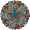
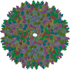
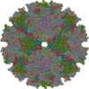
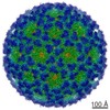
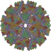
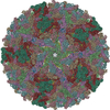
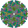
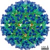
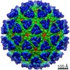
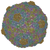
| Entry | Database: PDB / ID: 3j0f | ||||||
|---|---|---|---|---|---|---|---|
| Title | Sindbis virion | ||||||
 Components Components |
| ||||||
 Keywords Keywords | VIRUS / alphavirus / virus assembly / glycoprotein | ||||||
| Function / homology |  Function and homology information Function and homology informationicosahedral viral capsid, spike / togavirin / T=4 icosahedral viral capsid / host cell endoplasmic reticulum / ubiquitin-like protein ligase binding / channel activity / monoatomic ion transmembrane transport / clathrin-dependent endocytosis of virus by host cell / symbiont-mediated suppression of host toll-like receptor signaling pathway / membrane fusion ...icosahedral viral capsid, spike / togavirin / T=4 icosahedral viral capsid / host cell endoplasmic reticulum / ubiquitin-like protein ligase binding / channel activity / monoatomic ion transmembrane transport / clathrin-dependent endocytosis of virus by host cell / symbiont-mediated suppression of host toll-like receptor signaling pathway / membrane fusion / host cell Golgi apparatus / entry receptor-mediated virion attachment to host cell / serine-type endopeptidase activity / viral translational frameshifting / fusion of virus membrane with host endosome membrane / viral envelope / virion attachment to host cell / host cell nucleus / host cell plasma membrane / virion membrane / structural molecule activity / proteolysis / RNA binding Similarity search - Function | ||||||
| Biological species |  Sindbis virus Sindbis virus | ||||||
| Method | ELECTRON MICROSCOPY / single particle reconstruction / cryo EM / Resolution: 7 Å | ||||||
 Authors Authors | Tang, J. / Jose, J. / Zhang, W. / Chipman, P. / Kuhn, R.J. / Baker, T.S. | ||||||
 Citation Citation | Journal: J Mol Biol / Year: 2011 Title: Molecular links between the E2 envelope glycoprotein and nucleocapsid core in Sindbis virus. Authors: Jinghua Tang / Joyce Jose / Paul Chipman / Wei Zhang / Richard J Kuhn / Timothy S Baker /  Abstract: A three-dimensional reconstruction of Sindbis virus at 7.0 Å resolution presented here provides a detailed view of the virion structure and includes structural evidence for key interactions that ...A three-dimensional reconstruction of Sindbis virus at 7.0 Å resolution presented here provides a detailed view of the virion structure and includes structural evidence for key interactions that occur between the capsid protein (CP) and transmembrane (TM) glycoproteins E1 and E2. Based on crystal structures of component proteins and homology modeling, we constructed a nearly complete, pseudo-atomic model of the virus. Notably, this includes identification of the 33-residue cytoplasmic domain of E2 (cdE2), which follows a path from the E2 TM helix to the CP where it enters and exits the CP hydrophobic pocket and then folds back to contact the viral membrane. Modeling analysis identified three major contact regions between cdE2 and CP, and the roles of specific residues were probed by molecular genetics. This identified R393 and E395 of cdE2 and Y162 and K252 of CP as critical for virus assembly. The N-termini of the CPs form a contiguous network that interconnects 12 pentameric and 30 hexameric CP capsomers. A single glycoprotein spike cross-links three neighboring CP capsomers as might occur during initiation of virus budding. | ||||||
| History |
|
- Structure visualization
| Movie |
Movie viewer |
|---|---|
| Structure viewer | Molecule: MolmilJmol/JSmol |
- Downloads & links
-Download
| PDBx/mmCIF format | 3j0f.cif.gz | 139.5 KB | Display | PDBx/mmCIF format |
|---|---|---|---|---|
| PDB format | pdb3j0f.ent.gz | 90.6 KB | Display | PDB format |
| PDBx/mmJSON format | 3j0f.json.gz | Tree view | PDBx/mmJSON format | |
| Others |  Other downloads Other downloads |
-Validation report
| Arichive directory | https://data.pdbj.org/pub/pdb/validation_reports/j0/3j0fftp://data.pdbj.org/pub/pdb/validation_reports/j0/3j0f | HTTPS FTP |
|---|
-Related structure data
| Related structure data |  5251MC M: map data used to model this data C: citing same article ( |
|---|---|
| Similar structure data |
-Links
 PDBj
PDBj


- Assembly
| Deposited unit | 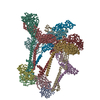
|
|---|---|
| 1 | x 60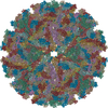
|
| 2 |
|
| 3 | x 5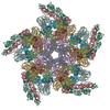
|
| 4 | x 6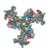
|
| 5 | 
|
| Symmetry | Point symmetry: (Schoenflies symbol: I (icosahedral)) |
-Components
| #1: Protein | Mass: 29410.010 Da / Num. of mol.: 4 / Fragment: UNP residues 1-264 / Source method: isolated from a natural source / Source: (natural) Sindbis virus / References: UniProt: P03316, togavirin#2: Protein | Mass: 47416.836 Da / Num. of mol.: 4 / Fragment: UNP residues 807-1245 / Source method: isolated from a natural source / Source: (natural) Sindbis virus / References: UniProt: P03316#3: Protein | Mass: 46982.582 Da / Num. of mol.: 4 / Fragment: UNP residues 329-751 / Source method: isolated from a natural source / Source: (natural) Sindbis virus / References: UniProt: P03316 |
|---|
-Experimental details
-Experiment
| Experiment | Method: ELECTRON MICROSCOPY |
|---|---|
| EM experiment | Aggregation state: PARTICLE / 3D reconstruction method: single particle reconstruction |
- Sample preparation
| Component | Name: sindbis virion / Type: VIRUS / Details: alphavirus |
|---|---|
| Details of virus | Empty: NO / Enveloped: YES / Host category: VERTEBRATES / Isolate: STRAIN / Type: VIRION |
| Natural host | Organism: Homo sapiens |
| Specimen | Embedding applied: NO / Shadowing applied: NO / Staining applied: NO / Vitrification applied: YES |
| Vitrification | Cryogen name: ETHANE |
- Electron microscopy imaging
| Microscopy | Model: FEI/PHILIPS CM300FEG/T / Date: Dec 12, 2001 |
|---|---|
| Electron gun | Electron source:  FIELD EMISSION GUN / Accelerating voltage: 300 kV / Illumination mode: FLOOD BEAM FIELD EMISSION GUN / Accelerating voltage: 300 kV / Illumination mode: FLOOD BEAM |
| Electron lens | Mode: BRIGHT FIELD / Camera length: 0 mm |
| Specimen holder | Specimen holder model: GATAN LIQUID NITROGEN / Specimen holder type: eucentric / Tilt angle max: 0 ° / Tilt angle min: 0 ° |
| Image recording | Film or detector model: KODAK SO-163 FILM |
| Radiation | Protocol: SINGLE WAVELENGTH / Monochromatic (M) / Laue (L): M / Scattering type: x-ray |
| Radiation wavelength | Relative weight: 1 |
- Processing
| Symmetry | Point symmetry: I (icosahedral) | ||||||||||||
|---|---|---|---|---|---|---|---|---|---|---|---|---|---|
| 3D reconstruction | Resolution: 7 Å / Symmetry type: POINT | ||||||||||||
| Atomic model building | Space: REAL | ||||||||||||
| Atomic model building | PDB-ID: 1KXF Pdb chain-ID: A / Accession code: 1KXF / Source name: PDB / Type: experimental model | ||||||||||||
| Refinement step | Cycle: LAST
|

