 Movie
Movie Controller

[English] 日本語
 Yorodumi
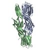
- PDB-2wvt: Crystal structure of an alpha-L-fucosidase GH29 from Bacteroides ... -
+ Open data

- Basic information
| Entry | Database: PDB / ID: 2wvt | ||||||
|---|---|---|---|---|---|---|---|
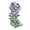
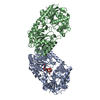
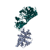
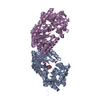
| Title | Crystal structure of an alpha-L-fucosidase GH29 from Bacteroides thetaiotaomicron in complex with a novel iminosugar fucosidase inhibitor | ||||||
 Components Components | ALPHA-L-FUCOSIDASE | ||||||
 Keywords Keywords | HYDROLASE / ALPHA-L-FUCOSE / GLYCOSIDE HYDROLASE FAMILY 29 | ||||||
| Function / homology |  Function and homology information Function and homology informationalpha-L-fucosidase / alpha-L-fucosidase activity / fucose metabolic process / glycoside catabolic process / lysosome Similarity search - Function | ||||||
| Biological species |  BACTEROIDES THETAIOTAOMICRON (bacteria) BACTEROIDES THETAIOTAOMICRON (bacteria) | ||||||
| Method |  X-RAY DIFFRACTION / SYNCHROTRON / MOLECULAR REPLACEMENT / Resolution: 1.8 Å X-RAY DIFFRACTION / SYNCHROTRON / MOLECULAR REPLACEMENT / Resolution: 1.8 Å | ||||||
 Authors Authors | Lammerts van Bueren, A. / Ardevol, A. / Fayers-Kerr, J. / Luo, B. / Zhang, Y. / Sollogoub, M. / Bleriot, Y. / Rovira, C. / Davies, G.J. | ||||||
 Citation Citation | Journal: J.Am.Chem.Soc. / Year: 2010 Title: Analysis of the Reaction Coordinate of Alpha-L-Fucosidases: A Combined Structural and Quantum Mechanical Approach. Authors: Lammerts Van Bueren, A. / Ardevol, A. / Fayers-Kerr, J. / Luo, B. / Zhang, Y. / Sollogoub, M. / Bleriot, Y. / Rovira, C. / Davies, G.J. | ||||||
| History |
|
- Structure visualization
| Structure viewer | Molecule: MolmilJmol/JSmol |
|---|
- Downloads & links
-Download
| PDBx/mmCIF format | 2wvt.cif.gz | 197.3 KB | Display | PDBx/mmCIF format |
|---|---|---|---|---|
| PDB format | pdb2wvt.ent.gz | 156.9 KB | Display | PDB format |
| PDBx/mmJSON format | 2wvt.json.gz | Tree view | PDBx/mmJSON format | |
| Others |  Other downloads Other downloads |
-Validation report
| Arichive directory | https://data.pdbj.org/pub/pdb/validation_reports/wv/2wvtftp://data.pdbj.org/pub/pdb/validation_reports/wv/2wvt | HTTPS FTP |
|---|
-Related structure data
| Related structure data |  2wvsC  2wvuC  2wvvSC C: citing same article ( S: Starting model for refinement |
|---|---|
| Similar structure data |


-Links
 PDBj
PDBj


- Assembly
| Deposited unit | 
| ||||||||
|---|---|---|---|---|---|---|---|---|---|
| 1 |
| ||||||||
| Unit cell |
|
-Components
| #1: Protein | Mass: 51331.121 Da / Num. of mol.: 2 / Fragment: RESIDUES 31-473 Source method: isolated from a genetically manipulated source Source: (gene. exp.) BACTEROIDES THETAIOTAOMICRON (bacteria)Strain: VPI-5482 / Plasmid: PET28 / Production host: #2: Chemical | 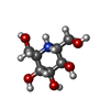  Mass: 194.206 Da / Num. of mol.: 2 / Source method: obtained synthetically / Formula: C7H16NO5 Mass: 194.206 Da / Num. of mol.: 2 / Source method: obtained synthetically / Formula: C7H16NO5#3: Chemical | ChemComp-GOL /   Mass: 92.094 Da / Num. of mol.: 5 / Source method: obtained synthetically / Formula: C3H8O3 Mass: 92.094 Da / Num. of mol.: 5 / Source method: obtained synthetically / Formula: C3H8O3#4: Chemical | ChemComp-IMD /   Mass: 69.085 Da / Num. of mol.: 4 / Source method: obtained synthetically / Formula: C3H5N2 Mass: 69.085 Da / Num. of mol.: 4 / Source method: obtained synthetically / Formula: C3H5N2#5: Water | ChemComp-HOH / |  Mass: 18.015 Da / Num. of mol.: 455 / Source method: isolated from a natural source / Formula: H2O Mass: 18.015 Da / Num. of mol.: 455 / Source method: isolated from a natural source / Formula: H2O |
|---|
-Experimental details
-Experiment
| Experiment | Method: X-RAY DIFFRACTION / Number of used crystals: 1 |
|---|
- Sample preparation
| Crystal | Density Matthews: 2.38 Å3/Da / Density % sol: 47.87 % / Description: NONE |
|---|
-Data collection
| Diffraction | Mean temperature: 120 K |
|---|---|
| Diffraction source | Source: SYNCHROTRON / Site: ESRF  / Beamline: ID23-1 / Wavelength: 0.99 / Beamline: ID23-1 / Wavelength: 0.99 |
| Detector | Type: ADSC CCD / Detector: CCD / Date: Apr 18, 2009 |
| Radiation | Protocol: SINGLE WAVELENGTH / Monochromatic (M) / Laue (L): M / Scattering type: x-ray |
| Radiation wavelength | Wavelength: 0.99 Å / Relative weight: 1 |
| Reflection | Resolution: 1.8→48 Å / Num. obs: 93456 / % possible obs: 99.9 % / Observed criterion σ(I): 2 / Redundancy: 3.7 % / Rmerge(I) obs: 0.1 / Net I/σ(I): 8.7 |
- Processing
| Software | Name: REFMAC / Version: 5.5.0102 / Classification: refinement | ||||||||||||||||||||||||||||||||||||||||||||||||||||||||||||||||||||||||||||||||||||||||||||||||||||||||||||||||||||||||||||||||||||||||||||||||||||||||||||||||||||||||||||||||||||||
|---|---|---|---|---|---|---|---|---|---|---|---|---|---|---|---|---|---|---|---|---|---|---|---|---|---|---|---|---|---|---|---|---|---|---|---|---|---|---|---|---|---|---|---|---|---|---|---|---|---|---|---|---|---|---|---|---|---|---|---|---|---|---|---|---|---|---|---|---|---|---|---|---|---|---|---|---|---|---|---|---|---|---|---|---|---|---|---|---|---|---|---|---|---|---|---|---|---|---|---|---|---|---|---|---|---|---|---|---|---|---|---|---|---|---|---|---|---|---|---|---|---|---|---|---|---|---|---|---|---|---|---|---|---|---|---|---|---|---|---|---|---|---|---|---|---|---|---|---|---|---|---|---|---|---|---|---|---|---|---|---|---|---|---|---|---|---|---|---|---|---|---|---|---|---|---|---|---|---|---|---|---|---|---|
| Refinement | Method to determine structure: MOLECULAR REPLACEMENT Starting model: PDB ENTRY 2WVV Resolution: 1.8→90.93 Å / Cor.coef. Fo:Fc: 0.948 / Cor.coef. Fo:Fc free: 0.926 / SU B: 3.471 / SU ML: 0.105 / Cross valid method: THROUGHOUT / ESU R: 0.138 / ESU R Free: 0.132 / Stereochemistry target values: MAXIMUM LIKELIHOOD Details: HYDROGENS HAVE BEEN ADDED IN THE RIDING POSITIONS. U VALUES REFINED INDIVIDUALLY
| ||||||||||||||||||||||||||||||||||||||||||||||||||||||||||||||||||||||||||||||||||||||||||||||||||||||||||||||||||||||||||||||||||||||||||||||||||||||||||||||||||||||||||||||||||||||
| Solvent computation | Ion probe radii: 0.8 Å / Shrinkage radii: 0.8 Å / VDW probe radii: 1.4 Å / Solvent model: MASK | ||||||||||||||||||||||||||||||||||||||||||||||||||||||||||||||||||||||||||||||||||||||||||||||||||||||||||||||||||||||||||||||||||||||||||||||||||||||||||||||||||||||||||||||||||||||
| Displacement parameters | Biso mean: 19.158 Å2
| ||||||||||||||||||||||||||||||||||||||||||||||||||||||||||||||||||||||||||||||||||||||||||||||||||||||||||||||||||||||||||||||||||||||||||||||||||||||||||||||||||||||||||||||||||||||
| Refinement step | Cycle: LAST / Resolution: 1.8→90.93 Å
| ||||||||||||||||||||||||||||||||||||||||||||||||||||||||||||||||||||||||||||||||||||||||||||||||||||||||||||||||||||||||||||||||||||||||||||||||||||||||||||||||||||||||||||||||||||||
| Refine LS restraints |
|