 Movie
Movie Controller

[English] 日本語
 Yorodumi
- PDB-2hhw: ddTTP:O6-methyl-guanine pair in the polymerase active site, in th... -
+ Open data

- Basic information
| Entry | Database: PDB / ID: 2hhw | ||||||||||||
|---|---|---|---|---|---|---|---|---|---|---|---|---|---|
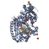
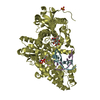
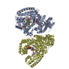
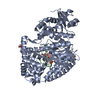
| Title | ddTTP:O6-methyl-guanine pair in the polymerase active site, in the closed conformation | ||||||||||||
 Components Components |
| ||||||||||||
 Keywords Keywords | Transferase/DNA / DNA polymerase I / DNA replication / Klenow fragment / DNA-protein complex / O6-methyl-guanine / Transferase-DNA COMPLEX | ||||||||||||
| Function / homology |  Function and homology information Function and homology information5'-3' exonuclease activity / 3'-5' exonuclease activity / DNA-templated DNA replication / double-strand break repair / DNA-directed DNA polymerase / DNA-directed DNA polymerase activity / nucleotide binding / DNA binding / metal ion binding Similarity search - Function | ||||||||||||
| Biological species |   Geobacillus stearothermophilus (bacteria) Geobacillus stearothermophilus (bacteria) | ||||||||||||
| Method |  X-RAY DIFFRACTION / SYNCHROTRON / MOLECULAR REPLACEMENT / Resolution: 1.88 Å X-RAY DIFFRACTION / SYNCHROTRON / MOLECULAR REPLACEMENT / Resolution: 1.88 Å | ||||||||||||
 Authors Authors | Warren, J.J. / Forsberg, L.J. / Beese, L.S. | ||||||||||||
 Citation Citation | Journal: Proc.Natl.Acad.Sci.Usa / Year: 2006 Title: The structural basis for the mutagenicity of O6-methyl-guanine lesions. Authors: Warren, J.J. / Forsberg, L.J. / Beese, L.S. | ||||||||||||
| History |
| ||||||||||||
| Remark 999 | SEQUENCE THE SEQUENCE OF THIS PROTEIN IS NOT AVAILABLE AT UNP SEQUENCE DATABASE AT THE TIME OF ...SEQUENCE THE SEQUENCE OF THIS PROTEIN IS NOT AVAILABLE AT UNP SEQUENCE DATABASE AT THE TIME OF PROCESSING. THE SEQUENCE OF THIS PROTEIN IS ANALOGOUS TO GEOBACILLUS KAUSTOPHILUS, UNP ACCESSION, Q5KWC1_GEOKA. |
- Structure visualization
| Structure viewer | Molecule: MolmilJmol/JSmol |
|---|
- Downloads & links
-Download
| PDBx/mmCIF format | 2hhw.cif.gz | 288.7 KB | Display | PDBx/mmCIF format |
|---|---|---|---|---|
| PDB format | pdb2hhw.ent.gz | 223.7 KB | Display | PDB format |
| PDBx/mmJSON format | 2hhw.json.gz | Tree view | PDBx/mmJSON format | |
| Others |  Other downloads Other downloads |
-Validation report
| Arichive directory | https://data.pdbj.org/pub/pdb/validation_reports/hh/2hhwftp://data.pdbj.org/pub/pdb/validation_reports/hh/2hhw | HTTPS FTP |
|---|
-Related structure data
| Related structure data |  2hhqC  2hhsC 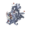 2hhtC  2hhuC  2hhvC  2hhxC  2hvhC  2hviC  2hw3C  1lv5S  2hhy C: citing same article ( S: Starting model for refinement |
|---|---|
| Similar structure data |


-Links
 PDBj
PDBj


- Assembly
| Deposited unit | 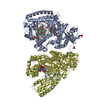
| ||||||||
|---|---|---|---|---|---|---|---|---|---|
| 1 | 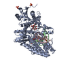
| ||||||||
| 2 | 
| ||||||||
| Unit cell |
| ||||||||
| Details | The asymmetric unit contains two biological assemblies, each consisting of one protein molecule, two strands of DNA, and an incoming dideoxy thymidine triphosphate. Chains A, B, C, and G constitute one assembly, and chains D, E, F, and G constitute the second. |
-Components
-DNA chain , 2 types, 4 molecules EBFC
| #1: DNA chain | Mass: 2675.775 Da / Num. of mol.: 2 / Source method: obtained synthetically / Details: chemically synthesized #2: DNA chain | Mass: 4030.650 Da / Num. of mol.: 2 / Source method: obtained synthetically / Details: chemically synthesized |
|---|
-Protein / Sugars , 2 types, 4 molecules AD
| #3: Protein | Mass: 66144.836 Da / Num. of mol.: 2 Fragment: residues 299-876 (analogous to E coli Klenow fragment) Mutation: D598A, F710Y Source method: isolated from a genetically manipulated source Source: (gene. exp.) Geobacillus stearothermophilus (bacteria)Gene: polA / Plasmid: PUC / Species (production host): Escherichia coli / Production host: #4: Polysaccharide |   Source method: isolated from a genetically manipulated source Details: oligosaccharide with reducing-end-to-reducing-end glycosidic bond References: sucrose |
|---|
-Non-polymers , 4 types, 801 molecules 


| #5: Chemical |  Mass: 54.938 Da / Num. of mol.: 2 / Source method: obtained synthetically / Formula: Mn Mass: 54.938 Da / Num. of mol.: 2 / Source method: obtained synthetically / Formula: Mn#6: Chemical |  Mass: 96.063 Da / Num. of mol.: 3 / Source method: obtained synthetically / Formula: SO4 Mass: 96.063 Da / Num. of mol.: 3 / Source method: obtained synthetically / Formula: SO4#7: Chemical |  Type: DNA OH 3 prime terminus / Mass: 466.169 Da / Num. of mol.: 2 / Source method: obtained synthetically / Formula: C10H17N2O13P3 Type: DNA OH 3 prime terminus / Mass: 466.169 Da / Num. of mol.: 2 / Source method: obtained synthetically / Formula: C10H17N2O13P3#8: Water | ChemComp-HOH / | Mass: 18.015 Da / Num. of mol.: 794 / Source method: isolated from a natural source / Formula: H2O |
|---|
-Experimental details
-Experiment
| Experiment | Method: X-RAY DIFFRACTION / Number of used crystals: 1 |
|---|
- Sample preparation
| Crystal | Density Matthews: 2.64 Å3/Da / Density % sol: 53.49 % | ||||||||||||||||||||||||||||||||
|---|---|---|---|---|---|---|---|---|---|---|---|---|---|---|---|---|---|---|---|---|---|---|---|---|---|---|---|---|---|---|---|---|---|
| Crystal grow | Temperature: 290 K / Method: vapor diffusion, hanging drop / pH: 5.8 Details: 50% saturated ammonium sulfate, 2.5% MPD, 100mM MES, pH 5.8, VAPOR DIFFUSION, HANGING DROP, temperature 290K | ||||||||||||||||||||||||||||||||
| Components of the solutions |
|
-Data collection
| Diffraction | Mean temperature: 100 K |
|---|---|
| Diffraction source | Source: SYNCHROTRON / Site: APS  / Beamline: 19-ID / Wavelength: 1.008 Å / Beamline: 19-ID / Wavelength: 1.008 Å |
| Detector | Type: CUSTOM-MADE / Detector: CCD / Date: Dec 14, 2004 |
| Radiation | Monochromator: Si 111 / Protocol: SINGLE WAVELENGTH / Monochromatic (M) / Laue (L): M / Scattering type: x-ray |
| Radiation wavelength | Wavelength: 1.008 Å / Relative weight: 1 |
| Reflection | Resolution: 1.88→50 Å / Num. all: 120016 / Num. obs: 120016 / % possible obs: 95.6 % / Observed criterion σ(I): -3 / Redundancy: 4.8 % / Rsym value: 0.111 / Χ2: 1.046 / Net I/σ(I): 14.2 |
| Reflection shell | Resolution: 1.88→1.95 Å / Redundancy: 4.4 % / Mean I/σ(I) obs: 2 / Num. unique all: 11633 / Rsym value: 0.782 / Χ2: 1.025 / % possible all: 93.6 |
- Processing
| Software |
| ||||||||||||||||||||||||||||
|---|---|---|---|---|---|---|---|---|---|---|---|---|---|---|---|---|---|---|---|---|---|---|---|---|---|---|---|---|---|
| Refinement | Method to determine structure: MOLECULAR REPLACEMENT Starting model: PDB entry 1LV5 Resolution: 1.88→50 Å / Isotropic thermal model: isotropic / σ(F): 0
| ||||||||||||||||||||||||||||
| Solvent computation | Bsol: 45.062 Å2 | ||||||||||||||||||||||||||||
| Displacement parameters | Biso mean: 24.212 Å2
| ||||||||||||||||||||||||||||
| Refine analyze |
| ||||||||||||||||||||||||||||
| Refinement step | Cycle: LAST / Resolution: 1.88→50 Å
| ||||||||||||||||||||||||||||
| Refine LS restraints |
| ||||||||||||||||||||||||||||
| LS refinement shell | Resolution: 1.88→1.95 Å
| ||||||||||||||||||||||||||||
| Xplor file |
|
