 Movie
Movie Controller

[English] 日本語
 Yorodumi
- PDB-3ez5: Cocrystal structure of Bacillus fragment DNA polymerase I with du... -
+ Open data

- Basic information
| Entry | Database: PDB / ID: 3ez5 | |||||||||
|---|---|---|---|---|---|---|---|---|---|---|
| Title | Cocrystal structure of Bacillus fragment DNA polymerase I with duplex DNA , dCTP, and zinc (closed form). | |||||||||
 Components Components |
| |||||||||
 Keywords Keywords | TRANSFERASE/DNA / protein-DNA complex / TRANSFERASE-DNA COMPLEX | |||||||||
| Function / homology |  Function and homology information Function and homology information5'-3' exonuclease activity / 3'-5' exonuclease activity / DNA-templated DNA replication / double-strand break repair / DNA-directed DNA polymerase / DNA-directed DNA polymerase activity / DNA binding Similarity search - Function | |||||||||
| Biological species |   Bacillus stearothermophilus (bacteria) Bacillus stearothermophilus (bacteria) | |||||||||
| Method |  X-RAY DIFFRACTION / SYNCHROTRON / Resolution: 1.9 Å X-RAY DIFFRACTION / SYNCHROTRON / Resolution: 1.9 Å | |||||||||
 Authors Authors | Warren, J.J. / Wu, E.Y. / Golosov, A.A. / Karplus, M. / Beese, L.S. | |||||||||
 Citation Citation | Journal: Structure / Year: 2010 Title: The Mechanism of the Translocation Step in DNA Replication by DNA Polymerase I: A Computer Simulation Analysis. Authors: Golosov, A.A. / Warren, J.J. / Beese, L.S. / Karplus, M. | |||||||||
| History |
|
- Structure visualization
| Structure viewer | Molecule: MolmilJmol/JSmol |
|---|
- Downloads & links
-Download
| PDBx/mmCIF format | 3ez5.cif.gz | 282.7 KB | Display | PDBx/mmCIF format |
|---|---|---|---|---|
| PDB format | pdb3ez5.ent.gz | 219.9 KB | Display | PDB format |
| PDBx/mmJSON format | 3ez5.json.gz | Tree view | PDBx/mmJSON format | |
| Others |  Other downloads Other downloads |
-Validation report
| Arichive directory | https://data.pdbj.org/pub/pdb/validation_reports/ez/3ez5ftp://data.pdbj.org/pub/pdb/validation_reports/ez/3ez5 | HTTPS FTP |
|---|
-Related structure data
| Related structure data |  3eyzC  2hviS C: citing same article ( S: Starting model for refinement |
|---|---|
| Similar structure data |


-Links
 PDBj
PDBj


- Assembly
| Deposited unit | 
| ||||||||
|---|---|---|---|---|---|---|---|---|---|
| 1 | 
| ||||||||
| 2 | 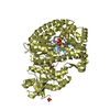
| ||||||||
| Unit cell |
|
-Components
-DNA chain , 2 types, 4 molecules BECF
| #2: DNA chain | Mass: 2691.774 Da / Num. of mol.: 2 / Source method: obtained synthetically / Details: DNA primer strand #3: DNA chain | Mass: 3702.428 Da / Num. of mol.: 2 / Source method: obtained synthetically / Details: DNA template strand |
|---|
-Protein / Sugars , 2 types, 4 molecules AD
| #1: Protein | Mass: 66202.945 Da / Num. of mol.: 2 / Mutation: F710Y Source method: isolated from a genetically manipulated source Details: The enzyme comes from a bacterium isolated from an Idaho hot spring, which was identified as a strain of Bacillus stearothermophilus by 16S rRNA sequence analysis (Kiefer, J.R. et al., 1997 Structure 5:95-108). Source: (gene. exp.) Bacillus stearothermophilus (bacteria) / Gene: POLA / Plasmid: pUC / Production host: References: UniProt: P52026*PLUS, DNA-directed DNA polymerase #4: Polysaccharide |   Source method: isolated from a genetically manipulated source Details: oligosaccharide with reducing-end-to-reducing-end glycosidic bond References: sucrose |
|---|
-Non-polymers , 4 types, 550 molecules 


| #5: Chemical |  Mass: 96.063 Da / Num. of mol.: 3 / Source method: obtained synthetically / Formula: SO4 Mass: 96.063 Da / Num. of mol.: 3 / Source method: obtained synthetically / Formula: SO4#6: Chemical | ChemComp-ZN /  Mass: 65.409 Da / Num. of mol.: 5 / Source method: obtained synthetically / Formula: Zn Mass: 65.409 Da / Num. of mol.: 5 / Source method: obtained synthetically / Formula: Zn#7: Chemical |  Mass: 475.182 Da / Num. of mol.: 2 / Source method: obtained synthetically / Formula: C10H16N5O11P3 Mass: 475.182 Da / Num. of mol.: 2 / Source method: obtained synthetically / Formula: C10H16N5O11P3#8: Water | ChemComp-HOH / | Mass: 18.015 Da / Num. of mol.: 540 / Source method: isolated from a natural source / Formula: H2O |
|---|
-Experimental details
-Experiment
| Experiment | Method: X-RAY DIFFRACTION / Number of used crystals: 1 |
|---|
- Sample preparation
| Crystal | Density Matthews: 2.59 Å3/Da / Density % sol: 52.53 % | ||||||||||||||||||||||||||||
|---|---|---|---|---|---|---|---|---|---|---|---|---|---|---|---|---|---|---|---|---|---|---|---|---|---|---|---|---|---|
| Crystal grow | Temperature: 290 K / Method: vapor diffusion, hanging drop / pH: 5.8 Details: Ammonium Sulfate, MES, MPD, pH 5.8, VAPOR DIFFUSION, HANGING DROP, temperature 290K | ||||||||||||||||||||||||||||
| Components of the solutions |
|
-Data collection
| Diffraction | Mean temperature: 100 K |
|---|---|
| Diffraction source | Source: SYNCHROTRON / Site: APS  / Beamline: 22-ID / Wavelength: 1.28242 Å / Beamline: 22-ID / Wavelength: 1.28242 Å |
| Detector | Type: MARMOSAIC 300 mm CCD / Detector: CCD / Date: Oct 21, 2004 |
| Radiation | Monochromator: Si 111 / Protocol: SINGLE WAVELENGTH / Monochromatic (M) / Laue (L): M / Scattering type: x-ray |
| Radiation wavelength | Wavelength: 1.28242 Å / Relative weight: 1 |
| Reflection | Resolution: 1.9→50 Å / Num. obs: 119138 / % possible obs: 99.9 % / Observed criterion σ(I): -3 / Redundancy: 3.78 % / Biso Wilson estimate: 39.668 Å2 / Rmerge(I) obs: 0.058 / Net I/σ(I): 12.28 |
| Reflection shell | Resolution: 1.9→2 Å / Redundancy: 3.7 % / Rmerge(I) obs: 0.398 / Mean I/σ(I) obs: 3.4 / Num. measured obs: 121167 / Num. unique all: 121167 / Num. unique obs: 32718 / % possible all: 100 |
- Processing
| Software |
| |||||||||||||||||||||||||||||||||||||||||||||||||||||||||||||||||
|---|---|---|---|---|---|---|---|---|---|---|---|---|---|---|---|---|---|---|---|---|---|---|---|---|---|---|---|---|---|---|---|---|---|---|---|---|---|---|---|---|---|---|---|---|---|---|---|---|---|---|---|---|---|---|---|---|---|---|---|---|---|---|---|---|---|---|
| Refinement | Starting model: PDB ENTRY 2HVI Resolution: 1.9→46.83 Å / Cor.coef. Fo:Fc: 0.953 / Cor.coef. Fo:Fc free: 0.934 / WRfactor Rfree: 0.278 / WRfactor Rwork: 0.235 / Occupancy max: 1 / Occupancy min: 0.2 / FOM work R set: 0.83 / SU B: 3.672 / SU ML: 0.109 / SU R Cruickshank DPI: 0.159 / SU Rfree: 0.145 / Isotropic thermal model: Isotropic / Cross valid method: THROUGHOUT / σ(F): -3 / ESU R: 0.159 / ESU R Free: 0.145 / Stereochemistry target values: MAXIMUM LIKELIHOOD Details: HYDROGENS HAVE BEEN ADDED IN THE RIDING POSITIONS U VALUES : REFINED INDIVIDUALLY
| |||||||||||||||||||||||||||||||||||||||||||||||||||||||||||||||||
| Solvent computation | Ion probe radii: 0.8 Å / Shrinkage radii: 0.8 Å / VDW probe radii: 1.2 Å / Solvent model: MASK | |||||||||||||||||||||||||||||||||||||||||||||||||||||||||||||||||
| Displacement parameters | Biso max: 89.23 Å2 / Biso mean: 37.225 Å2 / Biso min: 15.09 Å2
| |||||||||||||||||||||||||||||||||||||||||||||||||||||||||||||||||
| Refine analyze |
| |||||||||||||||||||||||||||||||||||||||||||||||||||||||||||||||||
| Refinement step | Cycle: LAST / Resolution: 1.9→46.83 Å
| |||||||||||||||||||||||||||||||||||||||||||||||||||||||||||||||||
| Refine LS restraints |
| |||||||||||||||||||||||||||||||||||||||||||||||||||||||||||||||||
| LS refinement shell | Resolution: 1.9→1.949 Å / Total num. of bins used: 20
|
