 Movie
Movie Controller


+ Open data

- Basic information
| Entry | Database: EMDB / ID: EMD-23543 | |||||||||
|---|---|---|---|---|---|---|---|---|---|---|
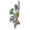
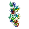
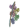
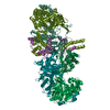
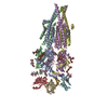
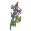
| Title | cryoEM structure DrdV-DNA complex | |||||||||
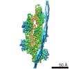
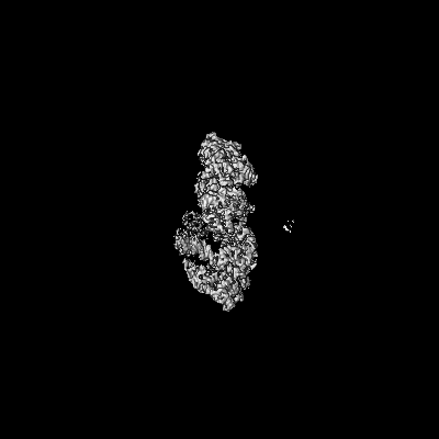
 Map data Map data | DrdVDNA complex dimer map from Glacios | |||||||||
 Sample Sample |
| |||||||||
 Keywords Keywords | inhibitor / Complex / endonuclease / methyl transferase / TypeIIL RM system / HYDROLASE / HYDROLASE-DNA complex | |||||||||
| Function / homology |  Function and homology information Function and homology informationN-methyltransferase activity / site-specific DNA-methyltransferase (adenine-specific) / site-specific DNA-methyltransferase (adenine-specific) activity / DNA restriction-modification system / endonuclease activity / methylation / DNA binding Similarity search - Function | |||||||||
| Biological species |  Deinococcus wulumuqiensis (bacteria) / synthetic construct (others) Deinococcus wulumuqiensis (bacteria) / synthetic construct (others) | |||||||||
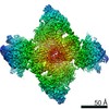
| Method | single particle reconstruction / cryo EM / Resolution: 3.25 Å | |||||||||
 Authors Authors | Shen BW / Stoddard BL | |||||||||
| Funding support |  United States, 1 items United States, 1 items
| |||||||||
 Citation Citation | Journal: Structure / Year: 2021 Title: Coordination of phage genome degradation versus host genome protection by a bifunctional restriction-modification enzyme visualized by CryoEM. Authors: Betty W Shen / Joel D Quispe / Yvette Luyten / Benjamin E McGough / Richard D Morgan / Barry L Stoddard / Abstract: Restriction enzymes that combine methylation and cleavage into a single assemblage and modify one DNA strand are capable of efficient adaptation toward novel targets. However, they must reliably ...Restriction enzymes that combine methylation and cleavage into a single assemblage and modify one DNA strand are capable of efficient adaptation toward novel targets. However, they must reliably cleave invasive DNA and methylate newly replicated unmodified host sites. One possible solution is to enforce a competition between slow methylation at a single unmodified host target, versus faster cleavage that requires multiple unmodified target sites in foreign DNA to be brought together in a reaction synapse. To examine this model, we have determined the catalytic behavior of a bifunctional type IIL restriction-modification enzyme and determined its structure, via cryoelectron microscopy, at several different stages of assembly and coordination with bound DNA targets. The structures demonstrate a mechanism in which an initial dimer is formed between two DNA-bound enzyme molecules, positioning the endonuclease domain from each enzyme against the other's DNA and requiring further additional DNA-bound enzyme molecules to enable cleavage. | |||||||||
| History |
|
- Structure visualization
| Movie |
Movie viewer |
|---|---|
| Structure viewer | EM map: SurfViewMolmilJmol/JSmol |
| Supplemental images |

- Downloads & links
-EMDB archive
| Map data | emd_23543.map.gz | 122.9 MB | EMDB map data format | |
|---|---|---|---|---|
| Header (meta data) | emd-23543-v30.xmlemd-23543.xml | 16.8 KB 16.8 KB | Display Display | EMDB header |
| Images | 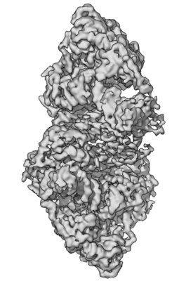 emd_23543.png emd_23543.png | 48.9 KB | ||
| Filedesc metadata | emd-23543.cif.gz | 6.8 KB | ||
| Archive directory |  http://ftp.pdbj.org/pub/emdb/structures/EMD-23543ftp://ftp.pdbj.org/pub/emdb/structures/EMD-23543 http://ftp.pdbj.org/pub/emdb/structures/EMD-23543ftp://ftp.pdbj.org/pub/emdb/structures/EMD-23543 | HTTPS FTP |
-Related structure data
| Related structure data | 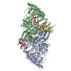 7lvvMC 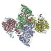 7lo5C M: atomic model generated by this map C: citing same article ( |
|---|---|
| Similar structure data |

-Links
| EMDB pages | EMDB (EBI/PDBe) / EMDataResource |
|---|
-Map
| File | Download / File: emd_23543.map.gz / Format: CCP4 / Size: 244.1 MB / Type: IMAGE STORED AS FLOATING POINT NUMBER (4 BYTES) | ||||||||||||||||||||||||||||||||||||||||||||||||||||||||||||
|---|---|---|---|---|---|---|---|---|---|---|---|---|---|---|---|---|---|---|---|---|---|---|---|---|---|---|---|---|---|---|---|---|---|---|---|---|---|---|---|---|---|---|---|---|---|---|---|---|---|---|---|---|---|---|---|---|---|---|---|---|---|
| Annotation | DrdVDNA complex dimer map from Glacios | ||||||||||||||||||||||||||||||||||||||||||||||||||||||||||||
| Projections & slices | Image control
Images are generated by Spider. | ||||||||||||||||||||||||||||||||||||||||||||||||||||||||||||
| Voxel size | X=Y=Z: 1.16 Å | ||||||||||||||||||||||||||||||||||||||||||||||||||||||||||||
| Density |
| ||||||||||||||||||||||||||||||||||||||||||||||||||||||||||||
| Symmetry | Space group: 1 | ||||||||||||||||||||||||||||||||||||||||||||||||||||||||||||
| Details | EMDB XML:
CCP4 map header:
| ||||||||||||||||||||||||||||||||||||||||||||||||||||||||||||
 Z (Sec.)
 Y (Row.)
 X (Col.)


-Supplemental data
- Sample components
-Entire : DrdVDNA complex
| Entire | Name: DrdVDNA complex |
|---|---|
| Components |
|
-Supramolecule #1: DrdVDNA complex
| Supramolecule | Name: DrdVDNA complex / type: complex / ID: 1 / Parent: 0 / Macromolecule list: #1-#3 / Details: dimer of DrdVDNA complex |
|---|---|
| Source (natural) | Organism: Deinococcus wulumuqiensis (bacteria) |
| Molecular weight | Theoretical: 0.24 kDa/nm |
-Macromolecule #1: Site-specific DNA-methyltransferase (adenine-specific)
| Macromolecule | Name: Site-specific DNA-methyltransferase (adenine-specific) type: protein_or_peptide / ID: 1 / Number of copies: 4 / Enantiomer: LEVO EC number: site-specific DNA-methyltransferase (adenine-specific) |
|---|---|
| Source (natural) | Organism: Deinococcus wulumuqiensis (bacteria) |
| Molecular weight | Theoretical: 118.256859 KDa |
| Recombinant expression | Organism: |
| Sequence | String: MSLQLVKKFQ KRLEDIVAYG GTRNESSVRA AFQQLLSDWA EGSGLRLITE VTQKAVAGNN VRPDGTLKDS LQQSRGYWES KDEADTLDD EIQKKLAKGY PRDNIIFEDS RLAVLMQNGE EVQRVDMGDA GALAGLLKLF FEFEPPQVLE FRKAVDHFKD E MPHLLKIL ...String: MSLQLVKKFQ KRLEDIVAYG GTRNESSVRA AFQQLLSDWA EGSGLRLITE VTQKAVAGNN VRPDGTLKDS LQQSRGYWES KDEADTLDD EIQKKLAKGY PRDNIIFEDS RLAVLMQNGE EVQRVDMGDA GALAGLLKLF FEFEPPQVLE FRKAVDHFKD E MPHLLKIL REAADAAEQK ADYRGERDHF VEIAKEAINP DFSPRDAREM LIQHILTGDL FTSVFDNAQY HEDNNIAQQL QQ LAATFYK GPVKRDIAER TKRYYGAIQA AAAQIADHHE KQRFLKALYE NFYRAYNPAG AERLGIFYTP GEIVRFMIEA TDT LLEKHF QKELADKGVE ILDPATGTGT FITELIDFLP KAKLEQKYRE ELHCNELALL PYYIANLNIE ATYAQKMGRY EEFR NIVLV DTLDNTGFGV HGQQSGLFGS VTAENLERAK RQNARPVRVI IGNPPYRANQ ANENDNNKNR EYKEIDRRIK ATYVA ASTA QKTKLYDMYS RFLRWATDRL KEDGIVAFVS NSSFIDSRTF DGFRKEVVKD FDHIYILDMK GNANTSGERR KREGGN VFN DQIKVGVAVY FLVRSAAGKR KSKDTKIWYH AVPDFWRARE KLEWLKTTKF EDIEFDHIRP DAKHNWLGQV DEENDWN EF LPVADKDTKQ AKGLGQERAI FKLYSLGVVT NRDEWVYSRA EDELADKVRY FIGRYNEIIK LPLGDLMSRN WEGDIKMT R ATIADAQSRK SYSLEKNSIV PSLYRPFDVL KMYFSKNLNE MQYQMPSIFP KGVGENVVIA LSGSPAAKPF QVLATDILP SLDLLEKTQC LPFYRYTMNG ERLNNITDYA LKAFQTHYAD TSISREDIFH YVYAVLHHPA YREKYALNLR QEFPRIPFYP EFGRWAAWG RELMALHIGF ESVAPYPLKR TDEPPKNDTP EALALAKKAR LKVQRDAAKQ PTGAVELDGL TTLAGIPAAA W AYKLGNRS ALEWVLERHK ETTPKDATIR EKFNTYRFAD HKERVIDLLA RVTTVSVETV RIVGEMPAET M UniProtKB: site-specific DNA-methyltransferase (adenine-specific) |
-Macromolecule #2: DNA (28-MER)
| Macromolecule | Name: DNA (28-MER) / type: dna / ID: 2 / Number of copies: 2 / Classification: DNA |
|---|---|
| Source (natural) | Organism: synthetic construct (others) |
| Molecular weight | Theoretical: 8.748646 KDa |
| Sequence | String: (DC)(DA)(DG)(DC)(DC)(DC)(DA)(DT)(DG)(DG) (DA)(DC)(DC)(DC)(DA)(DG)(DA)(DA)(DC)(DC) (DA)(DC)(DC)(DC)(DA)(DC)(DC)(DC)(DG) |
-Macromolecule #3: DNA (27-MER)
| Macromolecule | Name: DNA (27-MER) / type: dna / ID: 3 / Number of copies: 2 / Classification: DNA |
|---|---|
| Source (natural) | Organism: synthetic construct (others) |
| Molecular weight | Theoretical: 9.085788 KDa |
| Sequence | String: (DG)(DG)(DG)(DT)(DG)(DG)(DG)(DT)(DG)(DG) (DT)(DT)(DC)(DT)(DG)(DG)(DG)(DT)(DC)(DC) (DA)(DT)(DG)(DG)(DG)(DC)(DT)(DG)(DC) |
-Macromolecule #4: S-ADENOSYLMETHIONINE
| Macromolecule | Name: S-ADENOSYLMETHIONINE / type: ligand / ID: 4 / Number of copies: 2 / Formula: SAM |
|---|---|
| Molecular weight | Theoretical: 398.437 Da |
| Chemical component information |  ChemComp-SAM: |
-Macromolecule #5: CALCIUM ION
| Macromolecule | Name: CALCIUM ION / type: ligand / ID: 5 / Number of copies: 4 / Formula: CA |
|---|---|
| Molecular weight | Theoretical: 40.078 Da |
-Experimental details
-Structure determination
| Method | cryo EM |
|---|---|
 Processing Processing | single particle reconstruction |
| Aggregation state | particle |
-Sample preparation
| Concentration | 0.25 mg/mL | ||||||
|---|---|---|---|---|---|---|---|
| Buffer | pH: 7.5 / Component:
| ||||||
| Grid | Model: Quantifoil R1.2/1.3 / Material: COPPER / Mesh: 200 / Support film - Material: CARBON / Support film - topology: HOLEY / Pretreatment - Type: GLOW DISCHARGE / Pretreatment - Time: 30 sec. / Pretreatment - Pressure: 0.015 kPa | ||||||
| Vitrification | Cryogen name: ETHANE / Chamber humidity: 95 % / Chamber temperature: 298 K / Instrument: FEI VITROBOT MARK IV |
- Electron microscopy
| Microscope | TFS GLACIOS |
|---|---|
| Temperature | Min: 70.0 K / Max: 70.0 K |
| Specialist optics | Energy filter - Slit width: 20 eV |
| Image recording | Film or detector model: FEI CETA (4k x 4k) / Number grids imaged: 3 / Number real images: 1200 / Average exposure time: 2.0 sec. / Average electron dose: 40.0 e/Å2 Details: images were collected in movie-mode at 5 frames per second |
| Electron beam | Acceleration voltage: 200 kV / Electron source:  FIELD EMISSION GUN FIELD EMISSION GUN |
| Electron optics | C2 aperture diameter: 50.0 µm / Illumination mode: SPOT SCAN / Imaging mode: BRIGHT FIELD / Cs: 2.7 mm / Nominal defocus max: 2.3000000000000003 µm / Nominal defocus min: 0.8 µm / Nominal magnification: 37000 |
| Sample stage | Specimen holder model: FEI TITAN KRIOS AUTOGRID HOLDER / Cooling holder cryogen: NITROGEN |
