Movie
Movie Controller

[English] 日本語
 Yorodumi
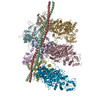
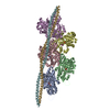
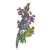
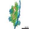
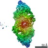
- PDB-3j4k: Cryo-EM structures of the actin:tropomyosin filament reveal the m... -
+ Open data

- Basic information
| Entry | Database: PDB / ID: 3j4k | ||||||
|---|---|---|---|---|---|---|---|
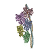
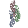
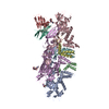
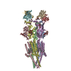
| Title | Cryo-EM structures of the actin:tropomyosin filament reveal the mechanism for the transition from C- to M-state | ||||||
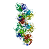
 Components Components |
| ||||||
 Keywords Keywords | STRUCTURAL PROTEIN / actin / tropomyosin / coiled-coil C-state | ||||||
| Function / homology |  Function and homology information Function and homology informationcytoskeletal motor activator activity / myosin heavy chain binding / tropomyosin binding / actin filament bundle / troponin I binding / filamentous actin / mesenchyme migration / skeletal muscle myofibril / actin filament bundle assembly / striated muscle thin filament ...cytoskeletal motor activator activity / myosin heavy chain binding / tropomyosin binding / actin filament bundle / troponin I binding / filamentous actin / mesenchyme migration / skeletal muscle myofibril / actin filament bundle assembly / striated muscle thin filament / skeletal muscle thin filament assembly / actin monomer binding / skeletal muscle fiber development / stress fiber / titin binding / actin filament polymerization / filopodium / actin filament / Hydrolases; Acting on acid anhydrides; Acting on acid anhydrides to facilitate cellular and subcellular movement / calcium-dependent protein binding / lamellipodium / cell body / protein domain specific binding / hydrolase activity / calcium ion binding / positive regulation of gene expression / magnesium ion binding / ATP binding / identical protein binding / cytoplasm Similarity search - Function | ||||||
| Biological species |   | ||||||
| Method | ELECTRON MICROSCOPY / single particle reconstruction / cryo EM / Resolution: 8 Å | ||||||
 Authors Authors | Sousa, D.R. / Stagg, S.M. / Stroupe, M.E. | ||||||
 Citation Citation | Journal: J Mol Biol / Year: 2013 Title: Cryo-EM structures of the actin:tropomyosin filament reveal the mechanism for the transition from C- to M-state. Authors: Duncan R Sousa / Scott M Stagg / M Elizabeth Stroupe /  Abstract: Tropomyosin (Tm) is a key factor in the molecular mechanisms that regulate the binding of myosin motors to actin filaments (F-Actins) in most eukaryotic cells. This regulation is achieved by the ...Tropomyosin (Tm) is a key factor in the molecular mechanisms that regulate the binding of myosin motors to actin filaments (F-Actins) in most eukaryotic cells. This regulation is achieved by the azimuthal repositioning of Tm along the actin (Ac):Tm:troponin (Tn) thin filament to block or expose myosin binding sites on Ac. In striated muscle, including involuntary cardiac muscle, Tm regulates muscle contraction by coupling Ca(2+) binding to Tn with myosin binding to the thin filament. In smooth muscle, the switch is the posttranslational modification of the myosin. Depending on the activation state of Tn and the binding state of myosin, Tm can occupy the blocked, closed, or open position on Ac. Using native cryogenic 3DEM (three-dimensional electron microscopy), we have directly resolved and visualized cardiac and gizzard muscle Tm on filamentous Ac in the position that corresponds to the closed state. From the 8-Å-resolution structure of the reconstituted Ac:Tm filament formed with gizzard-derived Tm, we discuss two possible mechanisms for the transition from closed to open state and describe the role Tm plays in blocking myosin tight binding in the closed-state position. | ||||||
| History |
|
- Structure visualization
| Movie |
Movie viewer |
|---|---|
| Structure viewer | Molecule: MolmilJmol/JSmol |
- Downloads & links
-Download
| PDBx/mmCIF format | 3j4k.cif.gz | 351 KB | Display | PDBx/mmCIF format |
|---|---|---|---|---|
| PDB format | pdb3j4k.ent.gz | 276.5 KB | Display | PDB format |
| PDBx/mmJSON format | 3j4k.json.gz | Tree view | PDBx/mmJSON format | |
| Others |  Other downloads Other downloads |
-Validation report
| Arichive directory | https://data.pdbj.org/pub/pdb/validation_reports/j4/3j4kftp://data.pdbj.org/pub/pdb/validation_reports/j4/3j4k | HTTPS FTP |
|---|
-Related structure data
| Related structure data | 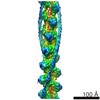 5751MC  5752C M: map data used to model this data C: citing same article ( |
|---|---|
| Similar structure data |
-Links
 PDBj
PDBj


- Assembly
| Deposited unit | 
|
|---|---|
| 1 |
|
-Components
| #1: Protein | Mass: 41862.613 Da / Num. of mol.: 5 / Source method: isolated from a natural source / Source: (natural) #2: Protein | Mass: 11592.281 Da / Num. of mol.: 2 / Source method: isolated from a natural source / Source: (natural) #3: Chemical | ChemComp-ADP /   Mass: 427.201 Da / Num. of mol.: 5 / Source method: obtained synthetically / Formula: C10H15N5O10P2 / Comment: ADP, energy-carrying molecule*YM Mass: 427.201 Da / Num. of mol.: 5 / Source method: obtained synthetically / Formula: C10H15N5O10P2 / Comment: ADP, energy-carrying molecule*YM |
|---|
-Experimental details
-Experiment
| Experiment | Method: ELECTRON MICROSCOPY |
|---|---|
| EM experiment | Aggregation state: FILAMENT / 3D reconstruction method: single particle reconstruction |
- Sample preparation
| Component | Name: F-actin/tropomyosin / Type: COMPLEX |
|---|---|
| Buffer solution | Name: 70 mM NaCl, 3 mM MgCl2, 0.2 mM EGTA, 5 mM NaH2PO4, 5 mM PIPES buffer pH: 7.5 Details: 70 mM NaCl, 3 mM MgCl2, 0.2 mM EGTA, 5 mM NaH2PO4, 5 mM PIPES buffer |
| Specimen | Embedding applied: NO / Shadowing applied: NO / Staining applied: NO / Vitrification applied: YES |
| Specimen support | Details: glow discharged Quantifoil 2/2 perforated carbon copper grids |
| Vitrification | Instrument: FEI VITROBOT MARK IV / Cryogen name: ETHANE / Humidity: 100 % / Chamber temperature: 293 K Details: 3 second blot before plunging into liquid ethane (FEI Vitrobot Mark IV) Method: 3 second blot |
- Electron microscopy imaging
| Experimental equipment |  Model: Titan Krios / Image courtesy: FEI Company |
|---|---|
| Microscopy | Model: FEI TITAN KRIOS / Date: Mar 2, 2010 |
| Electron gun | Electron source:  FIELD EMISSION GUN / Accelerating voltage: 300 kV / Illumination mode: FLOOD BEAM FIELD EMISSION GUN / Accelerating voltage: 300 kV / Illumination mode: FLOOD BEAM |
| Electron lens | Mode: BRIGHT FIELD / Nominal magnification: 59000 X / Calibrated magnification: 101555 X / Nominal defocus max: 4000 nm / Nominal defocus min: 1500 nm / Cs: 2 mm |
| Specimen holder | Temperature: 90 K / Tilt angle max: 0 ° / Tilt angle min: 0 ° |
| Image recording | Electron dose: 30 e/Å2 / Film or detector model: GATAN ULTRASCAN 4000 (4k x 4k) |
- Processing
| EM software |
| ||||||||||||||||||||
|---|---|---|---|---|---|---|---|---|---|---|---|---|---|---|---|---|---|---|---|---|---|
| CTF correction | Details: individual images | ||||||||||||||||||||
| Helical symmerty | Angular rotation/subunit: 167 ° / Axial rise/subunit: 28 Å / Axial symmetry: C1 | ||||||||||||||||||||
| 3D reconstruction | Method: IHRSR / Resolution: 8 Å / Resolution method: FSC 0.5 CUT-OFF / Num. of particles: 224337 / Nominal pixel size: 1.5 Å / Actual pixel size: 1.477 Å / Magnification calibration: helical diffraction / Symmetry type: HELICAL | ||||||||||||||||||||
| Atomic model building | Protocol: RIGID BODY FIT / Space: REAL / Target criteria: Cross correlation Details: REFINEMENT PROTOCOL--RIGID BODY DETAILS--The F-actin and tropomyosin were fitted separately. | ||||||||||||||||||||
| Atomic model building | 3D fitting-ID: 1 / Accession code: 4A7F / Initial refinement model-ID: 1 / PDB-ID: 4A7F / Source name: PDB / Type: experimental model
| ||||||||||||||||||||
| Refinement step | Cycle: LAST
|