Movie
Movie Controller


+ Open data

- Basic information
| Entry | Database: EMDB / ID: EMD-5751 | |||||||||
|---|---|---|---|---|---|---|---|---|---|---|
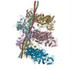
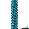
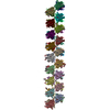
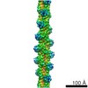
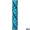
| Title | Cryogenic electron microscopy of cardiac actin:tropomyosin | |||||||||
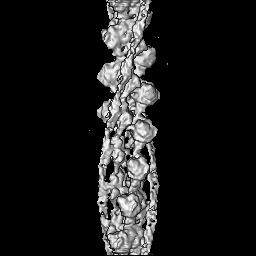
 Map data Map data | Reconstruction of rabbit skeletal muscle F-actin with chicken gizzard (smooth) muscle tropomyosin. A B-factor of -487 Angstrom^2 and an 8-Angstrom low pass filter were applied. | |||||||||
 Sample Sample |
| |||||||||
 Keywords Keywords | coiled-coil / actin-binding protein / cytoskeleton | |||||||||
| Function / homology |  Function and homology information Function and homology informationSmooth Muscle Contraction / muscle thin filament tropomyosin / Striated Muscle Contraction / cytoskeletal motor activator activity / myosin heavy chain binding / tropomyosin binding / actin filament bundle / troponin I binding / filamentous actin / mesenchyme migration ...Smooth Muscle Contraction / muscle thin filament tropomyosin / Striated Muscle Contraction / cytoskeletal motor activator activity / myosin heavy chain binding / tropomyosin binding / actin filament bundle / troponin I binding / filamentous actin / mesenchyme migration / skeletal muscle myofibril / actin filament bundle assembly / striated muscle thin filament / skeletal muscle thin filament assembly / actin monomer binding / skeletal muscle fiber development / stress fiber / titin binding / actin filament polymerization / muscle contraction / actin filament organization / filopodium / actin filament / Hydrolases; Acting on acid anhydrides; Acting on acid anhydrides to facilitate cellular and subcellular movement / calcium-dependent protein binding / actin filament binding / lamellipodium / actin cytoskeleton / cell body / protein heterodimerization activity / protein domain specific binding / hydrolase activity / calcium ion binding / positive regulation of gene expression / magnesium ion binding / protein homodimerization activity / ATP binding / identical protein binding / cytoplasm Similarity search - Function | |||||||||
| Biological species |   | |||||||||
| Method | single particle reconstruction / cryo EM / Resolution: 8.0 Å | |||||||||
 Authors Authors | Sousa DR / Stagg SM / Stroupe ME | |||||||||
 Citation Citation | Journal: J Mol Biol / Year: 2013 Title: Cryo-EM structures of the actin:tropomyosin filament reveal the mechanism for the transition from C- to M-state. Authors: Duncan R Sousa / Scott M Stagg / M Elizabeth Stroupe /  Abstract: Tropomyosin (Tm) is a key factor in the molecular mechanisms that regulate the binding of myosin motors to actin filaments (F-Actins) in most eukaryotic cells. This regulation is achieved by the ...Tropomyosin (Tm) is a key factor in the molecular mechanisms that regulate the binding of myosin motors to actin filaments (F-Actins) in most eukaryotic cells. This regulation is achieved by the azimuthal repositioning of Tm along the actin (Ac):Tm:troponin (Tn) thin filament to block or expose myosin binding sites on Ac. In striated muscle, including involuntary cardiac muscle, Tm regulates muscle contraction by coupling Ca(2+) binding to Tn with myosin binding to the thin filament. In smooth muscle, the switch is the posttranslational modification of the myosin. Depending on the activation state of Tn and the binding state of myosin, Tm can occupy the blocked, closed, or open position on Ac. Using native cryogenic 3DEM (three-dimensional electron microscopy), we have directly resolved and visualized cardiac and gizzard muscle Tm on filamentous Ac in the position that corresponds to the closed state. From the 8-Å-resolution structure of the reconstituted Ac:Tm filament formed with gizzard-derived Tm, we discuss two possible mechanisms for the transition from closed to open state and describe the role Tm plays in blocking myosin tight binding in the closed-state position. | |||||||||
| History |
|
- Structure visualization
| Movie |
Movie viewer |
|---|---|
| Structure viewer | EM map: SurfViewMolmilJmol/JSmol |
| Supplemental images |

- Downloads & links
-EMDB archive
| Map data | emd_5751.map.gz | 1.2 MB | EMDB map data format | |
|---|---|---|---|---|
| Header (meta data) | emd-5751-v30.xmlemd-5751.xml | 12.5 KB 12.5 KB | Display Display | EMDB header |
| Images | 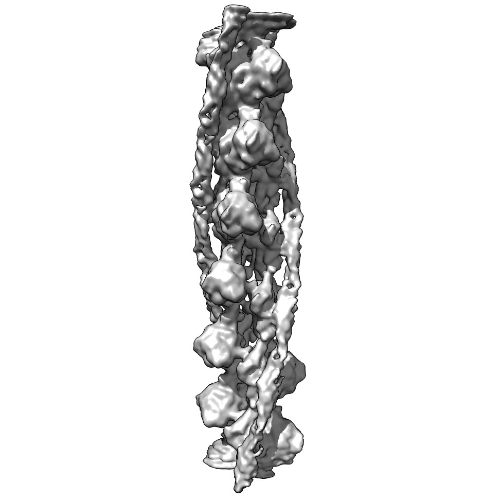 emd_5751_1.jpg emd_5751_1.jpg | 77.3 KB | ||
| Archive directory |  http://ftp.pdbj.org/pub/emdb/structures/EMD-5751ftp://ftp.pdbj.org/pub/emdb/structures/EMD-5751 http://ftp.pdbj.org/pub/emdb/structures/EMD-5751ftp://ftp.pdbj.org/pub/emdb/structures/EMD-5751 | HTTPS FTP |
-Related structure data
| Related structure data | 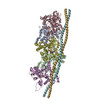 3j4kMC  5752C M: atomic model generated by this map C: citing same article ( |
|---|---|
| Similar structure data |

-Links
| EMDB pages | EMDB (EBI/PDBe) / EMDataResource |
|---|---|
| Related items in Molecule of the Month |


-Map
| File | Download / File: emd_5751.map.gz / Format: CCP4 / Size: 62.5 MB / Type: IMAGE STORED AS FLOATING POINT NUMBER (4 BYTES) | ||||||||||||||||||||||||||||||||||||||||||||||||||||||||||||
|---|---|---|---|---|---|---|---|---|---|---|---|---|---|---|---|---|---|---|---|---|---|---|---|---|---|---|---|---|---|---|---|---|---|---|---|---|---|---|---|---|---|---|---|---|---|---|---|---|---|---|---|---|---|---|---|---|---|---|---|---|---|
| Annotation | Reconstruction of rabbit skeletal muscle F-actin with chicken gizzard (smooth) muscle tropomyosin. A B-factor of -487 Angstrom^2 and an 8-Angstrom low pass filter were applied. | ||||||||||||||||||||||||||||||||||||||||||||||||||||||||||||
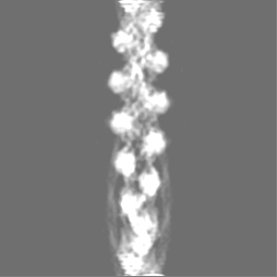
| Projections & slices | Image control
Images are generated by Spider. | ||||||||||||||||||||||||||||||||||||||||||||||||||||||||||||
| Voxel size | X=Y=Z: 1.477 Å | ||||||||||||||||||||||||||||||||||||||||||||||||||||||||||||
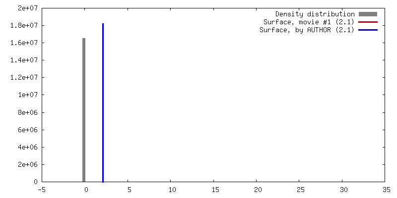
| Density |
| ||||||||||||||||||||||||||||||||||||||||||||||||||||||||||||
| Symmetry | Space group: 1 | ||||||||||||||||||||||||||||||||||||||||||||||||||||||||||||
| Details | EMDB XML:
CCP4 map header:
| ||||||||||||||||||||||||||||||||||||||||||||||||||||||||||||
 Z (Sec.)
 Y (Row.)
 X (Col.)


-Supplemental data
- Sample components
-Entire : rabbit skeletal F-actin bound to chicken gizzard tropomyosin
| Entire | Name: rabbit skeletal F-actin bound to chicken gizzard tropomyosin |
|---|---|
| Components |
|
-Supramolecule #1000: rabbit skeletal F-actin bound to chicken gizzard tropomyosin
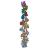
| Supramolecule | Name: rabbit skeletal F-actin bound to chicken gizzard tropomyosin type: sample / ID: 1000 Oligomeric state: fourteen actin monomers bind to two tropomyosin coiled-coils Number unique components: 2 |
|---|
-Macromolecule #1: Actin, alpha skeletal muscle
| Macromolecule | Name: Actin, alpha skeletal muscle / type: protein_or_peptide / ID: 1 / Name.synonym: Alpha-actin-1 Details: F-actin filaments were formed and tropomyosin was bound to the filaments. Oligomeric state: filament / Recombinant expression: No / Database: NCBI |
|---|---|
| Source (natural) | Organism: |
| Molecular weight | Experimental: 728 KDa |
| Sequence | UniProtKB: Actin, alpha skeletal muscle / GO: skeletal muscle thin filament assembly / InterPro: Actin family |
-Macromolecule #2: Tropomyosin alpha-1
| Macromolecule | Name: Tropomyosin alpha-1 / type: protein_or_peptide / ID: 2 / Name.synonym: Alpha-tropomyosin, Tropomyosin-1 Details: F-actin filaments were formed and tropomyosin was bound to the filaments. Oligomeric state: filament / Recombinant expression: No / Database: NCBI |
|---|---|
| Source (natural) | Organism: |
| Molecular weight | Experimental: 728 KDa |
| Sequence | UniProtKB: Tropomyosin beta chain / GO: muscle thin filament tropomyosin / InterPro: Tropomyosin |
-Experimental details
-Structure determination
| Method | cryo EM |
|---|---|
 Processing Processing | single particle reconstruction |
| Aggregation state | filament |
-Sample preparation
| Concentration | 0.5 mg/mL |
|---|---|
| Buffer | pH: 7.1 Details: 70 mM NaCl, 3 mM MgCl2, 0.2 mM EGTA, 5 mM NaH2PO4, 5 mM PIPES buffer |
| Grid | Details: Quantifoil 2/2 sized holes |
| Vitrification | Cryogen name: ETHANE / Chamber humidity: 100 % / Chamber temperature: 120 K / Instrument: FEI VITROBOT MARK IV Method: Blot with calcium-free filter paper for 2.5 seconds before plunging. |
- Electron microscopy
| Microscope | FEI TITAN KRIOS |
|---|---|
| Date | Mar 2, 2011 |
| Image recording | Category: CCD / Film or detector model: GATAN ULTRASCAN 4000 (4k x 4k) / Number real images: 700 / Average electron dose: 30 e/Å2 Details: Images are recorded on a Gatan Ultrascan 4000 CCD and are available upon request to MES (mestroupe@bio.fsu.edu) |
| Electron beam | Acceleration voltage: 120 kV / Electron source:  FIELD EMISSION GUN FIELD EMISSION GUN |
| Electron optics | Calibrated magnification: 101555 / Illumination mode: FLOOD BEAM / Imaging mode: BRIGHT FIELD / Nominal defocus max: 4.0 µm / Nominal defocus min: 1.5 µm / Nominal magnification: 59000 |
| Sample stage | Specimen holder model: FEI TITAN KRIOS AUTOGRID HOLDER |
| Experimental equipment |  Model: Titan Krios / Image courtesy: FEI Company |
-Image processing
| Details | Filaments were selected by hand via points along each filament and then boxed at even intervals. |
|---|---|
| CTF correction | Details: Micrograph |
| Final reconstruction | Applied symmetry - Helical parameters - Δz: 27.5 Å Applied symmetry - Helical parameters - Δ&Phi: 166.6 ° Applied symmetry - Helical parameters - Axial symmetry: C1 (asymmetric) Algorithm: OTHER / Resolution.type: BY AUTHOR / Resolution: 8.0 Å / Resolution method: FSC 0.5 CUT-OFF / Software - Name: Matlab, SPIDER, EMAN / Number images used: 224337 |
-Atomic model buiding 1
| Initial model | PDB ID: Chain - #0 - Chain ID: A / Chain - #1 - Chain ID: B / Chain - #2 - Chain ID: D / Chain - #3 - Chain ID: E / Chain - #4 - Chain ID: F / Chain - #5 - Chain ID: H / Chain - #6 - Chain ID: I |
|---|---|
| Software | Name: Chimera Fit-in-Map |
| Details | The F-actin and tropomyosin were fitted separately. |
| Refinement | Space: REAL / Protocol: RIGID BODY FIT / Target criteria: Cross correlation |
| Output model | PDB-3j4k: |