 Movie
Movie Controller

[English] 日本語
 Yorodumi
- PDB-6sjj: A new modulated crystal structure of ANS complex of St John's wor... -
+ Open data

- Basic information
| Entry | Database: PDB / ID: 6sjj | ||||||
|---|---|---|---|---|---|---|---|
| Title | A new modulated crystal structure of ANS complex of St John's wort Hyp-1 protein with 36 protein molecules in the asymmetric unit of the supercell | ||||||
 Components Components | PR-10 protein | ||||||
 Keywords Keywords | PLANT PROTEIN / PLANT HORMONE BINDING / PHYTOHORMONE BINDING / CYTOKININ / PLANT DEFENSE / PATHOGENESIS-RELATED PROTEIN / PR-10 PROTEIN / HYPERICIN / DEPRESSION / PR-10 FOLD / HYDROPHOBIC CAVITY / GLYCINE-RICH LOOP / ANS DISPLACEMENT ASSAY (ADA) / COMMENSURATELY MODULATED SUPERSTRUCTURE / TETARTOHEDRAL TWINNING | ||||||
| Function / homology |  Function and homology information Function and homology informationabscisic acid binding / abscisic acid-activated signaling pathway / protein phosphatase inhibitor activity / defense response / signaling receptor activity / nucleus / cytoplasm Similarity search - Function | ||||||
| Biological species |  Hypericum perforatum (plant) Hypericum perforatum (plant) | ||||||
| Method |  X-RAY DIFFRACTION / SYNCHROTRON / MOLECULAR REPLACEMENT / Resolution: 2.3 Å X-RAY DIFFRACTION / SYNCHROTRON / MOLECULAR REPLACEMENT / Resolution: 2.3 Å | ||||||
 Authors Authors | Smietanska, J. / Sliwiak, J. / Gilski, M. / Dauter, Z. / Strzalka, R. / Wolny, J. / Jaskolski, M. | ||||||
 Citation Citation | Journal: Acta Crystallogr D Struct Biol / Year: 2020 Title: A new modulated crystal structure of the ANS complex of the St John's wort Hyp-1 protein with 36 protein molecules in the asymmetric unit of the supercell. Authors: Smietanska, J. / Sliwiak, J. / Gilski, M. / Dauter, Z. / Strzalka, R. / Wolny, J. / Jaskolski, M. | ||||||
| History |
|
- Structure visualization
| Structure viewer | Molecule: MolmilJmol/JSmol |
|---|
- Downloads & links
-Download
| PDBx/mmCIF format | 6sjj.cif.gz | 1.1 MB | Display | PDBx/mmCIF format |
|---|---|---|---|---|
| PDB format | pdb6sjj.ent.gz | 999.5 KB | Display | PDB format |
| PDBx/mmJSON format | 6sjj.json.gz | Tree view | PDBx/mmJSON format | |
| Others |  Other downloads Other downloads |
-Validation report
| Arichive directory | https://data.pdbj.org/pub/pdb/validation_reports/sj/6sjjftp://data.pdbj.org/pub/pdb/validation_reports/sj/6sjj | HTTPS FTP |
|---|
-Related structure data
| Related structure data |  4n3eS S: Starting model for refinement |
|---|---|
| Similar structure data |

-Links
 PDBj
PDBj


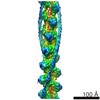
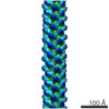
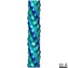
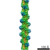
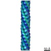
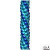
- Assembly
| Deposited unit | 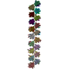
| ||||||||
|---|---|---|---|---|---|---|---|---|---|
| 1 |
| ||||||||
| Unit cell |
|
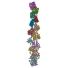
-Components
-Protein , 1 types, 36 molecules BARQPONMLKJIHGFEDCfedcbaZYXWVU...
| #1: Protein | Mass: 18324.898 Da / Num. of mol.: 36 / Mutation: 0 Source method: isolated from a genetically manipulated source Source: (gene. exp.) Hypericum perforatum (plant) / Gene: PR10.1 / Production host:  |
|---|
-Non-polymers , 6 types, 330 molecules 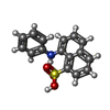


| #2: Chemical | ChemComp-2AN /  Mass: 299.344 Da / Num. of mol.: 157 / Source method: obtained synthetically / Formula: C16H13NO3S / Feature type: SUBJECT OF INVESTIGATION Mass: 299.344 Da / Num. of mol.: 157 / Source method: obtained synthetically / Formula: C16H13NO3S / Feature type: SUBJECT OF INVESTIGATION#3: Chemical | ChemComp-SO4 /  Mass: 96.063 Da / Num. of mol.: 10 / Source method: obtained synthetically / Formula: SO4 Mass: 96.063 Da / Num. of mol.: 10 / Source method: obtained synthetically / Formula: SO4#4: Chemical | ChemComp-EPE / |  Mass: 238.305 Da / Num. of mol.: 1 / Source method: obtained synthetically / Formula: C8H18N2O4S / Comment: pH buffer*YM Mass: 238.305 Da / Num. of mol.: 1 / Source method: obtained synthetically / Formula: C8H18N2O4S / Comment: pH buffer*YM#5: Chemical |  Mass: 78.133 Da / Num. of mol.: 3 / Source method: obtained synthetically / Formula: C2H6OS / Comment: DMSO, precipitant*YM Mass: 78.133 Da / Num. of mol.: 3 / Source method: obtained synthetically / Formula: C2H6OS / Comment: DMSO, precipitant*YM#6: Chemical | ChemComp-FLC /  Mass: 189.100 Da / Num. of mol.: 7 / Source method: obtained synthetically / Formula: C6H5O7 Mass: 189.100 Da / Num. of mol.: 7 / Source method: obtained synthetically / Formula: C6H5O7#7: Water | ChemComp-HOH / | Mass: 18.015 Da / Num. of mol.: 152 / Source method: isolated from a natural source / Formula: H2O |
|---|
-Details
| Has ligand of interest | Y |
|---|
-Experimental details
-Experiment
| Experiment | Method: X-RAY DIFFRACTION / Number of used crystals: 1 |
|---|
- Sample preparation
| Crystal | Density Matthews: 3.16 Å3/Da / Density % sol: 61.03 % |
|---|---|
| Crystal grow | Temperature: 292 K / Method: vapor diffusion, hanging drop Details: 0.1 M HEPES buffer pH 7.5, 1.3 M sodium citrate precipitant pH 6.3, 1:1 protein:reservoir volume ratio |
-Data collection
| Diffraction | Mean temperature: 100 K / Serial crystal experiment: N | |||||||||||||||||||||||||
|---|---|---|---|---|---|---|---|---|---|---|---|---|---|---|---|---|---|---|---|---|---|---|---|---|---|---|
| Diffraction source | Source: SYNCHROTRON / Site: BESSY  / Beamline: 14.2 / Wavelength: 0.918 Å / Beamline: 14.2 / Wavelength: 0.918 Å | |||||||||||||||||||||||||
| Detector | Type: DECTRIS PILATUS 2M / Detector: PIXEL / Date: Feb 12, 2014 | |||||||||||||||||||||||||
| Radiation | Monochromator: SI(111) crystal / Protocol: SINGLE WAVELENGTH / Monochromatic (M) / Laue (L): M / Scattering type: x-ray | |||||||||||||||||||||||||
| Radiation wavelength | Wavelength: 0.918 Å / Relative weight: 1 | |||||||||||||||||||||||||
| Reflection twin |
| |||||||||||||||||||||||||
| Reflection | Resolution: 2.3→31.58 Å / Num. obs: 127324 / % possible obs: 99.28 % / Redundancy: 7.2 % / Biso Wilson estimate: 67.46 Å2 / CC1/2: 0.978 / Rmerge(I) obs: 0.066 / Net I/σ(I): 6.22 | |||||||||||||||||||||||||
| Reflection shell | Resolution: 2.3→2.39 Å / Rmerge(I) obs: 0.691 / Num. unique obs: 24778 / CC1/2: 0.62 |
- Processing
| Software |
| ||||||||||||||||||||||||||||||||||||||||||||||||||||||||||||
|---|---|---|---|---|---|---|---|---|---|---|---|---|---|---|---|---|---|---|---|---|---|---|---|---|---|---|---|---|---|---|---|---|---|---|---|---|---|---|---|---|---|---|---|---|---|---|---|---|---|---|---|---|---|---|---|---|---|---|---|---|---|
| Refinement | Method to determine structure: MOLECULAR REPLACEMENT Starting model: 4N3E Resolution: 2.3→31.58 Å / Cor.coef. Fo:Fc: 0.965 / Cor.coef. Fo:Fc free: 0.953 / SU B: 7.176 / SU ML: 0.188 / Cross valid method: FREE R-VALUE / σ(F): 0 / ESU R: 0.062 / ESU R Free: 0.047 / Details: Hydrogen atoms were added at riding positions
| ||||||||||||||||||||||||||||||||||||||||||||||||||||||||||||
| Solvent computation | Ion probe radii: 0.8 Å / Shrinkage radii: 0.8 Å / VDW probe radii: 1.2 Å | ||||||||||||||||||||||||||||||||||||||||||||||||||||||||||||
| Displacement parameters | Biso max: 322.63 Å2 / Biso mean: 67.461 Å2 / Biso min: 16.02 Å2
| ||||||||||||||||||||||||||||||||||||||||||||||||||||||||||||
| Refinement step | Cycle: final / Resolution: 2.3→31.58 Å
| ||||||||||||||||||||||||||||||||||||||||||||||||||||||||||||
| Refine LS restraints |
| ||||||||||||||||||||||||||||||||||||||||||||||||||||||||||||
| LS refinement shell | Resolution: 2.301→2.356 Å / Rfactor Rfree error: 0
|
