 Movie
Movie Controller

[English] 日本語
 Yorodumi
- PDB-3f3p: Crystal structure of the nucleoporin pair Nup85-Seh1, space group... -
+ Open data

- Basic information
| Entry | Database: PDB / ID: 3f3p | ||||||
|---|---|---|---|---|---|---|---|
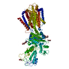
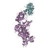
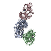
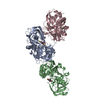
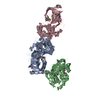
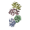
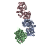
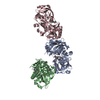
| Title | Crystal structure of the nucleoporin pair Nup85-Seh1, space group P21212 | ||||||
 Components Components |
| ||||||
 Keywords Keywords | STRUCTURAL PROTEIN / Protein Complex / Nucleoporin / Nucleoporin Complex / Nuclear Pore Complex / Macromolecular Assembly / Membrane Coat / Nucleocytoplasmic Transport / beta-propeller / solenoid domain / mRNA transport / Nucleus / Protein transport / Translocation / WD repeat | ||||||
| Function / homology |  Function and homology information Function and homology informationSeh1-associated complex / nuclear pore localization / regulation of TORC1 signaling / nuclear pore outer ring / Regulation of Glucokinase by Glucokinase Regulatory Protein / : / Regulation of HSF1-mediated heat shock response / structural constituent of nuclear pore / SUMOylation of SUMOylation proteins / SUMOylation of RNA binding proteins ...Seh1-associated complex / nuclear pore localization / regulation of TORC1 signaling / nuclear pore outer ring / Regulation of Glucokinase by Glucokinase Regulatory Protein / : / Regulation of HSF1-mediated heat shock response / structural constituent of nuclear pore / SUMOylation of SUMOylation proteins / SUMOylation of RNA binding proteins / SUMOylation of chromatin organization proteins / vacuolar membrane / nucleocytoplasmic transport / ribosomal large subunit export from nucleus / nuclear pore / mRNA transport / mRNA export from nucleus / positive regulation of TORC1 signaling / cellular response to amino acid starvation / protein import into nucleus / nuclear envelope / protein transport / nuclear membrane / positive regulation of DNA-templated transcription Similarity search - Function | ||||||
| Biological species |  | ||||||
| Method |  X-RAY DIFFRACTION / SYNCHROTRON / MOLECULAR REPLACEMENT / Resolution: 3.2 Å X-RAY DIFFRACTION / SYNCHROTRON / MOLECULAR REPLACEMENT / Resolution: 3.2 Å | ||||||
 Authors Authors | Debler, E.W. / Hseo, H. / Ma, Y. / Blobel, G. / Hoelz, A. | ||||||
 Citation Citation | Journal: Mol.Cell / Year: 2008 Title: A fence-like coat for the nuclear pore membrane. Authors: Debler, E.W. / Ma, Y. / Seo, H.S. / Hsia, K.C. / Noriega, T.R. / Blobel, G. / Hoelz, A. | ||||||
| History |
|
- Structure visualization
| Structure viewer | Molecule: MolmilJmol/JSmol |
|---|
- Downloads & links
-Download
| PDBx/mmCIF format | 3f3p.cif.gz | 926 KB | Display | PDBx/mmCIF format |
|---|---|---|---|---|
| PDB format | pdb3f3p.ent.gz | 760.6 KB | Display | PDB format |
| PDBx/mmJSON format | 3f3p.json.gz | Tree view | PDBx/mmJSON format | |
| Others |  Other downloads Other downloads |
-Validation report
| Arichive directory | https://data.pdbj.org/pub/pdb/validation_reports/f3/3f3pftp://data.pdbj.org/pub/pdb/validation_reports/f3/3f3p | HTTPS FTP |
|---|
-Related structure data
| Related structure data |  3f3fSC  3f3gC S: Starting model for refinement C: citing same article ( |
|---|---|
| Similar structure data |


-Links
 PDBj
PDBj

- Assembly
| Deposited unit | 
| ||||||||
|---|---|---|---|---|---|---|---|---|---|
| 1 | 
| ||||||||
| 2 | 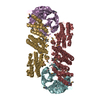
| ||||||||
| 3 | 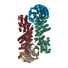
| ||||||||
| Unit cell |
| ||||||||
| Details | Authors state that the heterotetramers (ABCD), (EFGH), and (IJKL) constitute the biomolecules that build up the physiologically-relevant heterooctameric conformations represented by the asymmetric units of related entries 3F3F and 3F3G. |
-Components
| #1: Protein | Mass: 39640.488 Da / Num. of mol.: 6 Source method: isolated from a genetically manipulated source Source: (gene. exp.) Gene: SEH1 / Plasmid: pETDuet-1 / Production host:  #2: Protein | Mass: 65679.570 Da / Num. of mol.: 6 / Fragment: UNP residues 1-570 Source method: isolated from a genetically manipulated source Source: (gene. exp.) Gene: NUP85, RAT9 / Plasmid: pETDuet-1 / Production host: Has protein modification | Y | |
|---|
-Experimental details
-Experiment
| Experiment | Method: X-RAY DIFFRACTION / Number of used crystals: 1 |
|---|
- Sample preparation
| Crystal | Density Matthews: 3.62 Å3/Da / Density % sol: 66.06 % |
|---|---|
| Crystal grow | Temperature: 298 K / Method: vapor diffusion, hanging drop / pH: 6 Details: PEG 3350, Tacsimate pH 6.0, VAPOR DIFFUSION, HANGING DROP, temperature 298K |
-Data collection
| Diffraction | Mean temperature: 100 K |
|---|---|
| Diffraction source | Source: SYNCHROTRON / Site: APS  / Beamline: 23-ID-B / Wavelength: 1.0332 Å / Beamline: 23-ID-B / Wavelength: 1.0332 Å |
| Detector | Type: ADSC QUANTUM 315 / Detector: CCD / Date: Jul 4, 2008 |
| Radiation | Monochromator: double crystal / Protocol: SINGLE WAVELENGTH / Monochromatic (M) / Laue (L): M / Scattering type: x-ray |
| Radiation wavelength | Wavelength: 1.0332 Å / Relative weight: 1 |
| Reflection | Resolution: 3.2→50 Å / Num. all: 152129 / Num. obs: 150912 / % possible obs: 99.2 % / Observed criterion σ(I): -3 / Redundancy: 4.9 % / Rsym value: 0.145 / Net I/σ(I): 10.7 |
| Reflection shell | Resolution: 3.2→3.31 Å / Redundancy: 4.3 % / Num. unique all: 7406 / Rsym value: 0.729 / % possible all: 97.9 |
- Processing
| Software |
| ||||||||||||||||||||
|---|---|---|---|---|---|---|---|---|---|---|---|---|---|---|---|---|---|---|---|---|---|
| Refinement | Method to determine structure: MOLECULAR REPLACEMENT Starting model: PDB entry 3F3F Resolution: 3.2→50 Å / Stereochemistry target values: Engh & Huber
| ||||||||||||||||||||
| Refinement step | Cycle: LAST / Resolution: 3.2→50 Å
| ||||||||||||||||||||
| Refine LS restraints |
|
