 Movie
Movie Controller


+ Open data

- Basic information
| Entry | Database: PDB / ID: 6tu5 | ||||||
|---|---|---|---|---|---|---|---|
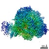
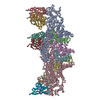
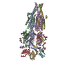
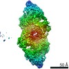
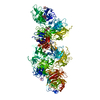
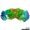
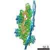
| Title | Influenza A/H7N9 polymerase core (apo) | ||||||
 Components Components |
| ||||||
 Keywords Keywords | VIRAL PROTEIN / RNA-dependent RNA polymerase | ||||||
| Function / homology |  Function and homology information Function and homology informationcap snatching / viral transcription / symbiont-mediated suppression of host mRNA transcription via inhibition of RNA polymerase II activity / host cell mitochondrion / 7-methylguanosine mRNA capping / virion component / endonuclease activity / Hydrolases; Acting on ester bonds / host cell cytoplasm / symbiont-mediated suppression of host gene expression ...cap snatching / viral transcription / symbiont-mediated suppression of host mRNA transcription via inhibition of RNA polymerase II activity / host cell mitochondrion / 7-methylguanosine mRNA capping / virion component / endonuclease activity / Hydrolases; Acting on ester bonds / host cell cytoplasm / symbiont-mediated suppression of host gene expression / viral translational frameshifting / RNA-directed RNA polymerase / viral RNA genome replication / nucleotide binding / hydrolase activity / RNA-directed RNA polymerase activity / host cell nucleus / DNA-templated transcription / RNA binding / metal ion binding Similarity search - Function | ||||||
| Biological species |   Influenza A virus Influenza A virus | ||||||
| Method |  X-RAY DIFFRACTION / SYNCHROTRON / MOLECULAR REPLACEMENT / Resolution: 3.325 Å X-RAY DIFFRACTION / SYNCHROTRON / MOLECULAR REPLACEMENT / Resolution: 3.325 Å | ||||||
 Authors Authors | Cusack, S. / Pflug, A. | ||||||
| Funding support |  France, 1items France, 1items
| ||||||
 Citation Citation | Journal: Cell / Year: 2020 Title: A Structure-Based Model for the Complete Transcription Cycle of Influenza Polymerase. Authors: Joanna M Wandzik / Tomas Kouba / Manikandan Karuppasamy / Alexander Pflug / Petra Drncova / Jan Provaznik / Nayara Azevedo / Stephen Cusack /  Abstract: Influenza polymerase uses unique mechanisms to synthesize capped and polyadenylated mRNAs from the genomic viral RNA (vRNA) template, which is packaged inside ribonucleoprotein particles (vRNPs). ...Influenza polymerase uses unique mechanisms to synthesize capped and polyadenylated mRNAs from the genomic viral RNA (vRNA) template, which is packaged inside ribonucleoprotein particles (vRNPs). Here, we visualize by cryoelectron microscopy the conformational dynamics of the polymerase during the complete transcription cycle from pre-initiation to termination, focusing on the template trajectory. After exiting the active site cavity, the template 3' extremity rebinds into a specific site on the polymerase surface. Here, it remains sequestered during all subsequent transcription steps, forcing the template to loop out as it further translocates. At termination, the strained connection between the bound template 5' end and the active site results in polyadenylation by stuttering at uridine 17. Upon product dissociation, further conformational changes release the trapped template, allowing recycling back into the pre-initiation state. Influenza polymerase thus performs transcription while tightly binding to and protecting both template ends, allowing efficient production of multiple mRNAs from a single vRNP. | ||||||
| History |
|
- Structure visualization
| Structure viewer | Molecule: MolmilJmol/JSmol |
|---|
- Downloads & links
-Download
| PDBx/mmCIF format | 6tu5.cif.gz | 554.2 KB | Display | PDBx/mmCIF format |
|---|---|---|---|---|
| PDB format | pdb6tu5.ent.gz | Display | PDB format | |
| PDBx/mmJSON format | 6tu5.json.gz | Tree view | PDBx/mmJSON format | |
| Others |  Other downloads Other downloads |
-Validation report
| Arichive directory | https://data.pdbj.org/pub/pdb/validation_reports/tu/6tu5ftp://data.pdbj.org/pub/pdb/validation_reports/tu/6tu5 | HTTPS FTP |
|---|
-Related structure data
| Related structure data |  6szuC 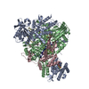 6szvC  6t0nC  6t0rC  6t0sC  6t0uC 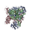 6t0vC 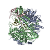 6t0wC  6t2cC  6tw1C  4wsbS S: Starting model for refinement C: citing same article ( |
|---|---|
| Similar structure data |
-Links
 PDBj
PDBj- Assembly
| Deposited unit | 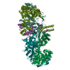
| ||||||||||||||||
|---|---|---|---|---|---|---|---|---|---|---|---|---|---|---|---|---|---|
| 1 |
| ||||||||||||||||
| Unit cell |
| ||||||||||||||||
| Noncrystallographic symmetry (NCS) | NCS domain:
NCS ensembles :
|
-Components
| #1: Protein | Mass: 59383.859 Da / Num. of mol.: 2 Source method: isolated from a genetically manipulated source Details: N-terminal truncation deletes endonuclease Source: (gene. exp.) Influenza A virus (A/Zhejiang/DTID-ZJU01/2013(H7N9))Strain: A/Zhejiang/DTID-ZJU01/2013(H7N9) / Gene: PA / Variant: H7N9 / Production host:  Trichoplusia ni (cabbage looper) Trichoplusia ni (cabbage looper)References: UniProt: M9TI86, Hydrolases; Acting on ester bonds #2: Protein | Mass: 86496.156 Da / Num. of mol.: 2 Source method: isolated from a genetically manipulated source Source: (gene. exp.) Influenza A virus (A/Zhejiang/DTID-ZJU01/2013(H7N9))Strain: A/Zhejiang/DTID-ZJU01/2013(H7N9) / Gene: PB1 / Variant: H7N9 / Production host: Trichoplusia ni (cabbage looper) / References: UniProt: M9TLW3, RNA-directed RNA polymerase#3: Protein | Mass: 16203.751 Da / Num. of mol.: 2 Source method: isolated from a genetically manipulated source Details: C-terminal sequence GSGSENLYFQ is linker and residual TEV cleavage site after cleavage Source: (gene. exp.) Influenza A virus (A/Zhejiang/DTID-ZJU01/2013(H7N9))Strain: A/Zhejiang/DTID-ZJU01/2013(H7N9) / Gene: PB2 / Variant: H7N9 / Production host: Trichoplusia ni (cabbage looper) / References: UniProt: X5F427#4: Chemical |   Mass: 24.305 Da / Num. of mol.: 2 / Source method: obtained synthetically / Formula: Mg Mass: 24.305 Da / Num. of mol.: 2 / Source method: obtained synthetically / Formula: MgHas ligand of interest | N | |
|---|
-Experimental details
-Experiment
| Experiment | Method: X-RAY DIFFRACTION / Number of used crystals: 1 |
|---|
- Sample preparation
| Crystal | Density Matthews: 2.8 Å3/Da / Density % sol: 56.01 % |
|---|---|
| Crystal grow | Temperature: 281 K / Method: vapor diffusion, sitting drop / pH: 7 Details: Protein at about 9 mg/ml 0.1 M Tris pH 7.0, 8-13% PEG 8K, 0.2 M MgCl2, 0.1 M guanidine hydrochloride |
-Data collection
| Diffraction | Mean temperature: 100 K / Serial crystal experiment: N |
|---|---|
| Diffraction source | Source: SYNCHROTRON / Site: ESRF / Beamline: MASSIF-3 / Wavelength: 0.9677 Å |
| Detector | Type: DECTRIS EIGER X 4M / Detector: PIXEL / Date: Jun 30, 2016 |
| Radiation | Protocol: SINGLE WAVELENGTH / Monochromatic (M) / Laue (L): M / Scattering type: x-ray |
| Radiation wavelength | Wavelength: 0.9677 Å / Relative weight: 1 |
| Reflection | Resolution: 3.325→132.024 Å / Num. obs: 50439 / % possible obs: 92.7 % / Redundancy: 11.6 % / CC1/2: 0.997 / Rmerge(I) obs: 0.19 / Rpim(I) all: 0.057 / Rrim(I) all: 0.199 / Net I/σ(I): 10.7 |
| Reflection shell | Resolution: 3.325→3.463 Å / Redundancy: 13.3 % / Rmerge(I) obs: 2.298 / Mean I/σ(I) obs: 1.5 / Num. unique obs: 2523 / CC1/2: 0.5 / Rrim(I) all: 2.388 / % possible all: 42.2 |
- Processing
| Software |
| ||||||||||||||||||||||||||||||||||||||||||||||||||||||||||||||||||||||||||||||||||||||||||||||||||||||||||||||||||||||||||||||||||||||||||||||||||||||||||||||||||||||||||
|---|---|---|---|---|---|---|---|---|---|---|---|---|---|---|---|---|---|---|---|---|---|---|---|---|---|---|---|---|---|---|---|---|---|---|---|---|---|---|---|---|---|---|---|---|---|---|---|---|---|---|---|---|---|---|---|---|---|---|---|---|---|---|---|---|---|---|---|---|---|---|---|---|---|---|---|---|---|---|---|---|---|---|---|---|---|---|---|---|---|---|---|---|---|---|---|---|---|---|---|---|---|---|---|---|---|---|---|---|---|---|---|---|---|---|---|---|---|---|---|---|---|---|---|---|---|---|---|---|---|---|---|---|---|---|---|---|---|---|---|---|---|---|---|---|---|---|---|---|---|---|---|---|---|---|---|---|---|---|---|---|---|---|---|---|---|---|---|---|---|---|---|
| Refinement | Method to determine structure: MOLECULAR REPLACEMENT Starting model: 4WSB Resolution: 3.325→132.024 Å / Cor.coef. Fo:Fc: 0.937 / Cor.coef. Fo:Fc free: 0.903 / SU B: 32.599 / SU ML: 0.499 / Cross valid method: FREE R-VALUE / ESU R Free: 0.617 Details: Hydrogens have been added in their riding positions
| ||||||||||||||||||||||||||||||||||||||||||||||||||||||||||||||||||||||||||||||||||||||||||||||||||||||||||||||||||||||||||||||||||||||||||||||||||||||||||||||||||||||||||
| Solvent computation | Ion probe radii: 0.8 Å / Shrinkage radii: 0.8 Å / VDW probe radii: 1.2 Å | ||||||||||||||||||||||||||||||||||||||||||||||||||||||||||||||||||||||||||||||||||||||||||||||||||||||||||||||||||||||||||||||||||||||||||||||||||||||||||||||||||||||||||
| Displacement parameters | Biso mean: 119.623 Å2
| ||||||||||||||||||||||||||||||||||||||||||||||||||||||||||||||||||||||||||||||||||||||||||||||||||||||||||||||||||||||||||||||||||||||||||||||||||||||||||||||||||||||||||
| Refinement step | Cycle: LAST / Resolution: 3.325→132.024 Å
| ||||||||||||||||||||||||||||||||||||||||||||||||||||||||||||||||||||||||||||||||||||||||||||||||||||||||||||||||||||||||||||||||||||||||||||||||||||||||||||||||||||||||||
| Refine LS restraints |
| ||||||||||||||||||||||||||||||||||||||||||||||||||||||||||||||||||||||||||||||||||||||||||||||||||||||||||||||||||||||||||||||||||||||||||||||||||||||||||||||||||||||||||
| LS refinement shell |
|
