 Movie
Movie Controller


+ Open data

- Basic information
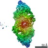
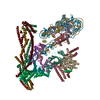
| Entry | Database: PDB / ID: 7lo5 | ||||||
|---|---|---|---|---|---|---|---|
| Title | cryoEM structure DrdV-DNA complex | ||||||
 Components Components |
| ||||||
 Keywords Keywords | HYDROLASE/DNA / inhibitor / Complex / endonuclease / methyl transferase / TypeIIL RM system / HYDROLASE / HYDROLASE-DNA complex | ||||||
| Function / homology |  Function and homology information Function and homology informationN-methyltransferase activity / site-specific DNA-methyltransferase (adenine-specific) / site-specific DNA-methyltransferase (adenine-specific) activity / DNA restriction-modification system / endonuclease activity / methylation / DNA binding Similarity search - Function | ||||||
| Biological species |  Deinococcus wulumuqiensis (bacteria) Deinococcus wulumuqiensis (bacteria)synthetic construct (others) | ||||||
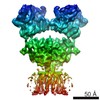
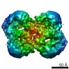
| Method | ELECTRON MICROSCOPY / single particle reconstruction / cryo EM / Resolution: 2.86 Å | ||||||
 Authors Authors | Shen, B.W. / Stoddard, B.L. | ||||||
| Funding support |  United States, 1items United States, 1items
| ||||||
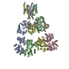
 Citation Citation | Journal: Structure / Year: 2021 Title: Coordination of phage genome degradation versus host genome protection by a bifunctional restriction-modification enzyme visualized by CryoEM. Authors: Betty W Shen / Joel D Quispe / Yvette Luyten / Benjamin E McGough / Richard D Morgan / Barry L Stoddard / Abstract: Restriction enzymes that combine methylation and cleavage into a single assemblage and modify one DNA strand are capable of efficient adaptation toward novel targets. However, they must reliably ...Restriction enzymes that combine methylation and cleavage into a single assemblage and modify one DNA strand are capable of efficient adaptation toward novel targets. However, they must reliably cleave invasive DNA and methylate newly replicated unmodified host sites. One possible solution is to enforce a competition between slow methylation at a single unmodified host target, versus faster cleavage that requires multiple unmodified target sites in foreign DNA to be brought together in a reaction synapse. To examine this model, we have determined the catalytic behavior of a bifunctional type IIL restriction-modification enzyme and determined its structure, via cryoelectron microscopy, at several different stages of assembly and coordination with bound DNA targets. The structures demonstrate a mechanism in which an initial dimer is formed between two DNA-bound enzyme molecules, positioning the endonuclease domain from each enzyme against the other's DNA and requiring further additional DNA-bound enzyme molecules to enable cleavage. | ||||||
| History |
|
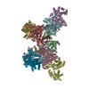
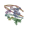
- Structure visualization
| Movie |
Movie viewer |
|---|---|
| Structure viewer | Molecule: MolmilJmol/JSmol |
- Downloads & links
-Download
| PDBx/mmCIF format | 7lo5.cif.gz | 818.1 KB | Display | PDBx/mmCIF format |
|---|---|---|---|---|
| PDB format | pdb7lo5.ent.gz | 664.2 KB | Display | PDB format |
| PDBx/mmJSON format | 7lo5.json.gz | Tree view | PDBx/mmJSON format | |
| Others |  Other downloads Other downloads |
-Validation report
| Arichive directory | https://data.pdbj.org/pub/pdb/validation_reports/lo/7lo5ftp://data.pdbj.org/pub/pdb/validation_reports/lo/7lo5 | HTTPS FTP |
|---|
-Related structure data
| Related structure data | 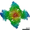 23461MC 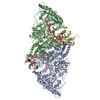 7lvvC M: map data used to model this data C: citing same article ( |
|---|---|
| Similar structure data |

-Links
 PDBj
PDBj


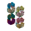
- Assembly
| Deposited unit | 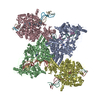
|
|---|---|
| 1 |
|
-Components
| #1: Protein | Mass: 118256.859 Da / Num. of mol.: 4 Source method: isolated from a genetically manipulated source Source: (gene. exp.) Deinococcus wulumuqiensis (bacteria) / Gene: drdVRM, DVJ83_12125 / Production host: References: UniProt: A0A345IJ72, site-specific DNA-methyltransferase (adenine-specific) #2: DNA chain | Mass: 8748.646 Da / Num. of mol.: 4 / Source method: obtained synthetically / Source: (synth.) synthetic construct (others) #3: DNA chain | Mass: 9085.788 Da / Num. of mol.: 4 / Source method: obtained synthetically / Source: (synth.) synthetic construct (others) #4: Chemical | ChemComp-SAM /   Mass: 398.437 Da / Num. of mol.: 4 / Source method: obtained synthetically / Formula: C15H22N6O5S / Feature type: SUBJECT OF INVESTIGATION Mass: 398.437 Da / Num. of mol.: 4 / Source method: obtained synthetically / Formula: C15H22N6O5S / Feature type: SUBJECT OF INVESTIGATION#5: Chemical | ChemComp-CA /   Mass: 40.078 Da / Num. of mol.: 4 / Source method: obtained synthetically / Formula: Ca / Feature type: SUBJECT OF INVESTIGATION Mass: 40.078 Da / Num. of mol.: 4 / Source method: obtained synthetically / Formula: Ca / Feature type: SUBJECT OF INVESTIGATIONHas ligand of interest | Y | |
|---|
-Experimental details
-Experiment
| Experiment | Method: ELECTRON MICROSCOPY |
|---|---|
| EM experiment | Aggregation state: PARTICLE / 3D reconstruction method: single particle reconstruction |
- Sample preparation
| Component | Name: DrdV-DNA tetramer II / Type: COMPLEX Details: DrdV-DNA complex after SEC Biorad 650 fractionation Entity ID: #1-#3 / Source: RECOMBINANT |
|---|---|
| Molecular weight | Value: 0.45 MDa / Experimental value: YES |
| Source (natural) | Organism: Deinococcus wulumuqiensis 479 (bacteria) |
| Source (recombinant) | Organism: |
| Buffer solution | pH: 8 / Details: 20 mM TrismaHCl ph 8.0/150 NaCl/2CaCl2 |
| Specimen | Conc.: 0.4 mg/ml / Embedding applied: NO / Shadowing applied: NO / Staining applied: NO / Vitrification applied: YES |
| Specimen support | Grid material: COPPER / Grid mesh size: 200 divisions/in. / Grid type: Quantifoil |
| Vitrification | Instrument: FEI VITROBOT MARK IV / Cryogen name: ETHANE / Humidity: 95 % / Chamber temperature: 298 K |
- Electron microscopy imaging
| Experimental equipment |  Model: Titan Krios / Image courtesy: FEI Company |
|---|---|
| Microscopy | Model: FEI TITAN KRIOS |
| Electron gun | Electron source:  FIELD EMISSION GUN / Accelerating voltage: 300 kV / Illumination mode: SPOT SCAN FIELD EMISSION GUN / Accelerating voltage: 300 kV / Illumination mode: SPOT SCAN |
| Electron lens | Mode: BRIGHT FIELD / Nominal defocus max: 2500 nm / Nominal defocus min: 1200 nm / Calibrated defocus min: 800 nm / Calibrated defocus max: 500 nm / Cs: 2.7 mm / C2 aperture diameter: 100 µm / Alignment procedure: COMA FREE |
| Specimen holder | Cryogen: NITROGEN / Specimen holder model: FEI TITAN KRIOS AUTOGRID HOLDER / Temperature (max): 70 K / Temperature (min): 70 K |
| Image recording | Average exposure time: 2 sec. / Electron dose: 30 e/Å2 / Film or detector model: GATAN K3 (6k x 4k) / Num. of grids imaged: 1 / Num. of real images: 4300 |
| EM imaging optics | Energyfilter name: GIF Quantum LS Chromatic aberration corrector: CEOS manufactured Cc corrector Energyfilter slit width: 20 eV Spherical aberration corrector: Cs corrector with two hexapod elements |
- Processing
| EM software |
| ||||||||||||||||||||||||||||||||||||||||
|---|---|---|---|---|---|---|---|---|---|---|---|---|---|---|---|---|---|---|---|---|---|---|---|---|---|---|---|---|---|---|---|---|---|---|---|---|---|---|---|---|---|
| CTF correction | Type: NONE | ||||||||||||||||||||||||||||||||||||||||
| Symmetry | Point symmetry: C1 (asymmetric) | ||||||||||||||||||||||||||||||||||||||||
| 3D reconstruction | Resolution: 2.86 Å / Resolution method: FSC 0.143 CUT-OFF / Num. of particles: 94920 / Algorithm: FOURIER SPACE / Num. of class averages: 4 / Symmetry type: POINT |
