[English] 日本語
Yorodumi
- EMDB-22293: Distinct conformational states of SARS-CoV-2 spike protein -

+
Open data


ID or keywords:

Loading...

-
Basic information

Entry
Database: EMDB / ID: EMD-22293
TitleDistinct conformational states of SARS-CoV-2 spike protein
Map datatop masked map
Sample
  • Complex: post-fusion SARS-CoV-2 spike glycoprotein
    • Protein or peptide: Spike glycoproteinSpike protein
  • Ligand: 2-acetamido-2-deoxy-beta-D-glucopyranose
  • Ligand: alpha-D-mannopyranose
Function / homology
Function and homology information


Maturation of spike protein / viral translation / Translation of Structural Proteins / Virion Assembly and Release / host cell surface / host extracellular space / suppression by virus of host tetherin activity / Induction of Cell-Cell Fusion / structural constituent of virion / host cell endoplasmic reticulum-Golgi intermediate compartment membrane ...Maturation of spike protein / viral translation / Translation of Structural Proteins / Virion Assembly and Release / host cell surface / host extracellular space / suppression by virus of host tetherin activity / Induction of Cell-Cell Fusion / structural constituent of virion / host cell endoplasmic reticulum-Golgi intermediate compartment membrane / entry receptor-mediated virion attachment to host cell / receptor-mediated endocytosis of virus by host cell / Attachment and Entry / membrane fusion / positive regulation of viral entry into host cell / receptor-mediated virion attachment to host cell / receptor ligand activity / host cell surface receptor binding / fusion of virus membrane with host plasma membrane / fusion of virus membrane with host endosome membrane / viral envelope / symbiont-mediated suppression of host type I interferon-mediated signaling pathway / virion attachment to host cell / SARS-CoV-2 activates/modulates innate and adaptive immune responses / host cell plasma membrane / virion membrane / membrane / identical protein binding / plasma membrane
Similarity search - Function
Spike (S) protein S1 subunit, receptor-binding domain, SARS-CoV-2 / Spike (S) protein S1 subunit, N-terminal domain, SARS-CoV-like / Betacoronavirus spike (S) glycoprotein S1 subunit N-terminal (NTD) domain profile. / Spike glycoprotein, N-terminal domain superfamily / Betacoronavirus spike (S) glycoprotein S1 subunit C-terminal (CTD) domain profile. / Spike glycoprotein, betacoronavirus / Spike (S) protein S1 subunit, receptor-binding domain, betacoronavirus / Spike S1 subunit, receptor binding domain superfamily, betacoronavirus / Betacoronavirus spike glycoprotein S1, receptor binding / Spike glycoprotein S1, N-terminal domain, betacoronavirus-like ...Spike (S) protein S1 subunit, receptor-binding domain, SARS-CoV-2 / Spike (S) protein S1 subunit, N-terminal domain, SARS-CoV-like / Betacoronavirus spike (S) glycoprotein S1 subunit N-terminal (NTD) domain profile. / Spike glycoprotein, N-terminal domain superfamily / Betacoronavirus spike (S) glycoprotein S1 subunit C-terminal (CTD) domain profile. / Spike glycoprotein, betacoronavirus / Spike (S) protein S1 subunit, receptor-binding domain, betacoronavirus / Spike S1 subunit, receptor binding domain superfamily, betacoronavirus / Betacoronavirus spike glycoprotein S1, receptor binding / Spike glycoprotein S1, N-terminal domain, betacoronavirus-like / Betacoronavirus-like spike glycoprotein S1, N-terminal / Spike glycoprotein S2, coronavirus, heptad repeat 1 / Spike glycoprotein S2, coronavirus, heptad repeat 2 / Coronavirus spike (S) glycoprotein S2 subunit heptad repeat 2 (HR2) region profile. / Coronavirus spike (S) glycoprotein S2 subunit heptad repeat 1 (HR1) region profile. / Spike glycoprotein S2 superfamily, coronavirus / Spike glycoprotein S2, coronavirus / Coronavirus spike glycoprotein S2 / Coronavirus spike glycoprotein S1, C-terminal / Coronavirus spike glycoprotein S1, C-terminal
Similarity search - Domain/homology
Biological speciesSevere acute respiratory syndrome coronavirus 2
Methodsingle particle reconstruction / cryo EM / Resolution: 3.0 Å
AuthorsZhang J / Cai YF / Xiao TS / Peng HQ / Sterling SM / Walsh Jr RM / Rawson S / Rits-Volloch S / Chen B
Funding support United States, 2 items
OrganizationGrant numberCountry
National Institutes of Health/National Institute Of Allergy and Infectious Diseases (NIH/NIAID)AI147884 United States
National Institutes of Health/National Institute Of Allergy and Infectious Diseases (NIH/NIAID)AI147884 01A1S1 United States
Citation
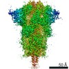
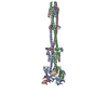
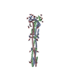
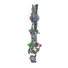
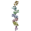
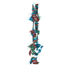
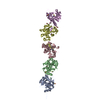
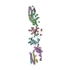
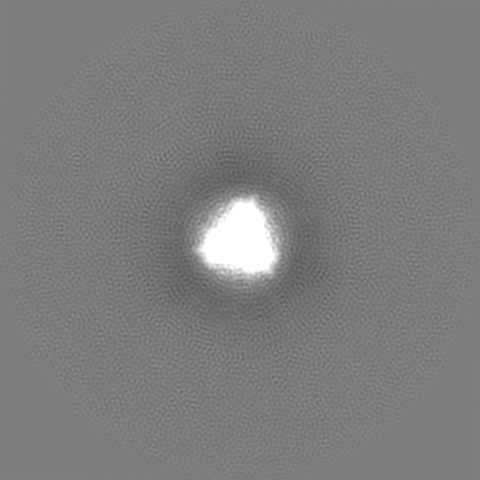
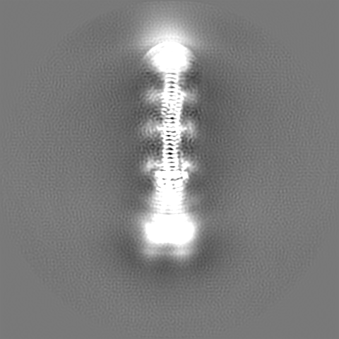
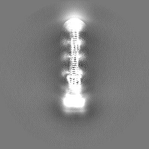
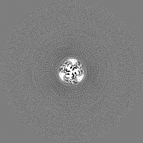
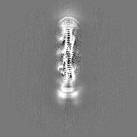
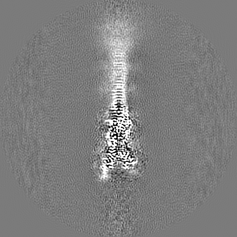
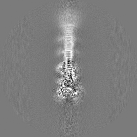
Journal: Science / Year: 2020
Title: Distinct conformational states of SARS-CoV-2 spike protein.
Authors: Yongfei Cai / Jun Zhang / Tianshu Xiao / Hanqin Peng / Sarah M Sterling / Richard M Walsh / Shaun Rawson / Sophia Rits-Volloch / Bing Chen /
Abstract: Intervention strategies are urgently needed to control the severe acute respiratory syndrome coronavirus 2 (SARS-CoV-2) pandemic. The trimeric viral spike (S) protein catalyzes fusion between viral ...Intervention strategies are urgently needed to control the severe acute respiratory syndrome coronavirus 2 (SARS-CoV-2) pandemic. The trimeric viral spike (S) protein catalyzes fusion between viral and target cell membranes to initiate infection. Here, we report two cryo-electron microscopy structures derived from a preparation of the full-length S protein, representing its prefusion (2.9-angstrom resolution) and postfusion (3.0-angstrom resolution) conformations, respectively. The spontaneous transition to the postfusion state is independent of target cells. The prefusion trimer has three receptor-binding domains clamped down by a segment adjacent to the fusion peptide. The postfusion structure is strategically decorated by N-linked glycans, suggesting possible protective roles against host immune responses and harsh external conditions. These findings advance our understanding of SARS-CoV-2 entry and may guide the development of vaccines and therapeutics.
History
DepositionJul 11, 2020-
Header (metadata) releaseJul 22, 2020-
Map releaseJul 22, 2020-
UpdateOct 7, 2020-
Current statusOct 7, 2020Processing site: RCSB / Status: Released

-
Structure visualization
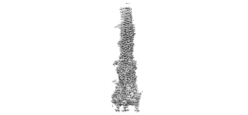
Movie
  • Surface view with section colored by density value
  • Surface level: 0.012
  • Imaged by UCSF Chimera
  • Download
  • Surface view colored by cylindrical radius
  • Surface level: 0.012
  • Imaged by UCSF Chimera
  • Download
  • Surface view with fitted model
  • Atomic models: PDB-6xra
  • Surface level: 0.012
  • Imaged by UCSF Chimera
  • Download
Movie viewer
Structure viewerEM map:
SurfViewMolmilJmol/JSmol
Supplemental images

Downloads & links

-
Map

FileDownload / File: emd_22293.map.gz / Format: CCP4 / Size: 421.9 MB / Type: IMAGE STORED AS FLOATING POINT NUMBER (4 BYTES)
Annotationtop masked map
Voxel sizeX=Y=Z: 0.825 Å
Density
Contour LevelBy AUTHOR: 0.007 / Movie #1: 0.012
Minimum - Maximum-0.072804056 - 0.1096557
Average (Standard dev.)-0.0000029053 (±0.0012398663)
SymmetrySpace group: 1
Details

EMDB XML:

Map geometry
Axis orderXYZ
Origin000
Dimensions480480480
Spacing480480480
CellA=B=C: 396.0 Å
α=β=γ: 90.0 °

CCP4 map header:

modeImage stored as Reals
Å/pix. X/Y/Z0.8250.8250.825
M x/y/z480480480
origin x/y/z0.0000.0000.000
length x/y/z396.000396.000396.000
α/β/γ90.00090.00090.000
start NX/NY/NZ434333
NX/NY/NZ116118137
MAP C/R/S123
start NC/NR/NS000
NC/NR/NS480480480
D min/max/mean-0.0730.110-0.000

-
Supplemental data

-
Additional map: bottom masked map

Fileemd_22293_additional_1.map
Annotationbottom masked map
Projections & Slices
AxesZYX

Projections

Slices (1/2)
Density Histograms

-
Additional map: top masked map

Fileemd_22293_additional_2.map
Annotationtop masked map
Projections & Slices
AxesZYX

Projections

Slices (1/2)
Density Histograms

-
Sample components

-
Entire : post-fusion SARS-CoV-2 spike glycoprotein

EntireName: post-fusion SARS-CoV-2 spike glycoprotein
Components
  • Complex: post-fusion SARS-CoV-2 spike glycoprotein
    • Protein or peptide: Spike glycoproteinSpike protein
  • Ligand: 2-acetamido-2-deoxy-beta-D-glucopyranose
  • Ligand: alpha-D-mannopyranose

-
Supramolecule #1: post-fusion SARS-CoV-2 spike glycoprotein

SupramoleculeName: post-fusion SARS-CoV-2 spike glycoprotein / type: complex / ID: 1 / Parent: 0 / Macromolecule list: #1
Details: overall map of post-fusion SARS-CoV-2 spike glycoprotein
Source (natural)Organism: Severe acute respiratory syndrome coronavirus 2
Recombinant expressionOrganism: Homo sapiens (human)
Molecular weightTheoretical: 210 kDa/nm

-
Macromolecule #1: Spike glycoprotein

MacromoleculeName: Spike glycoprotein / type: protein_or_peptide / ID: 1 / Number of copies: 3 / Enantiomer: LEVO
Source (natural)Organism: Severe acute respiratory syndrome coronavirus 2
Molecular weightTheoretical: 144.916062 KDa
Recombinant expressionOrganism: Homo sapiens (human)
SequenceString: MFVFLVLLPL VSSQCVNLTT RTQLPPAYTN SFTRGVYYPD KVFRSSVLHS TQDLFLPFFS NVTWFHAIHV SGTNGTKRFD NPVLPFNDG VYFASTEKSN IIRGWIFGTT LDSKTQSLLI VNNATNVVIK VCEFQFCNDP FLGVYYHKNN KSWMESEFRV Y SSANNCTF ...String:
MFVFLVLLPL VSSQCVNLTT RTQLPPAYTN SFTRGVYYPD KVFRSSVLHS TQDLFLPFFS NVTWFHAIHV SGTNGTKRFD NPVLPFNDG VYFASTEKSN IIRGWIFGTT LDSKTQSLLI VNNATNVVIK VCEFQFCNDP FLGVYYHKNN KSWMESEFRV Y SSANNCTF EYVSQPFLMD LEGKQGNFKN LREFVFKNID GYFKIYSKHT PINLVRDLPQ GFSALEPLVD LPIGINITRF QT LLALHRS YLTPGDSSSG WTAGAAAYYV GYLQPRTFLL KYNENGTITD AVDCALDPLS ETKCTLKSFT VEKGIYQTSN FRV QPTESI VRFPNITNLC PFGEVFNATR FASVYAWNRK RISNCVADYS VLYNSASFST FKCYGVSPTK LNDLCFTNVY ADSF VIRGD EVRQIAPGQT GKIADYNYKL PDDFTGCVIA WNSNNLDSKV GGNYNYLYRL FRKSNLKPFE RDISTEIYQA GSTPC NGVE GFNCYFPLQS YGFQPTNGVG YQPYRVVVLS FELLHAPATV CGPKKSTNLV KNKCVNFNFN GLTGTGVLTE SNKKFL PFQ QFGRDIADTT DAVRDPQTLE ILDITPCSFG GVSVITPGTN TSNQVAVLYQ DVNCTEVPVA IHADQLTPTW RVYSTGS NV FQTRAGCLIG AEHVNNSYEC DIPIGAGICA SYQTQTNSPR RARSVASQSI IAYTMSLGAE NSVAYSNNSI AIPTNFTI S VTTEILPVSM TKTSVDCTMY ICGDSTECSN LLLQYGSFCT QLNRALTGIA VEQDKNTQEV FAQVKQIYKT PPIKDFGGF NFSQILPDPS KPSKRSFIED LLFNKVTLAD AGFIKQYGDC LGDIAARDLI CAQKFNGLTV LPPLLTDEMI AQYTSALLAG TITSGWTFG AGAALQIPFA MQMAYRFNGI GVTQNVLYEN QKLIANQFNS AIGKIQDSLS STASALGKLQ DVVNQNAQAL N TLVKQLSS NFGAISSVLN DILSRLDKVE AEVQIDRLIT GRLQSLQTYV TQQLIRAAEI RASANLAATK MSECVLGQSK RV DFCGKGY HLMSFPQSAP HGVVFLHVTY VPAQEKNFTT APAICHDGKA HFPREGVFVS NGTHWFVTQR NFYEPQIITT DNT FVSGNC DVVIGIVNNT VYDPLQPELD SFKEELDKYF KNHTSPDVDL GDISGINASV VNIQKEIDRL NEVAKNLNES LIDL QELGK YEQYIKWPWY IWLGFIAGLI AIVMVTIMLC CMTSCCSCLK GCCSCGSCCK FDEDDSEPVL KGVKLHYTLE SGGGS AWSH PQFEKGGGSG GGSGGSSAWS HPQFEK

-
Macromolecule #5: 2-acetamido-2-deoxy-beta-D-glucopyranose

MacromoleculeName: 2-acetamido-2-deoxy-beta-D-glucopyranose / type: ligand / ID: 5 / Number of copies: 9 / Formula: NAG
Molecular weightTheoretical: 221.208 Da
Chemical component information

ChemComp-NAG:
2-acetamido-2-deoxy-beta-D-glucopyranose / N-Acetylglucosamine

-
Macromolecule #6: alpha-D-mannopyranose

MacromoleculeName: alpha-D-mannopyranose / type: ligand / ID: 6 / Number of copies: 3 / Formula: MAN
Molecular weightTheoretical: 180.156 Da
Chemical component information

ChemComp-MAN:
alpha-D-mannopyranose / Mannose

-
Experimental details

-
Structure determination

Methodcryo EM
Processingsingle particle reconstruction
Aggregation stateparticle

-
Sample preparation

Concentration0.3 mg/mL
BufferpH: 7.5
Component:
ConcentrationFormulaName
0.15 mol/lNaClSodium chloridesodium chloride
0.025 mol/lTris-HClTrisTris hydrochloride
0.02 %NP-40Nonidet P40
GridModel: Quantifoil / Material: GOLD / Support film - Material: CARBON / Support film - topology: CONTINUOUS / Support film - Film thickness: 0.2 nm
VitrificationCryogen name: ETHANE / Chamber humidity: 100 % / Chamber temperature: 277.15 K / Instrument: FEI VITROBOT MARK IV

-
Electron microscopy

MicroscopeFEI TITAN KRIOS
Electron beamAcceleration voltage: 300 kV / Electron source: FIELD EMISSION GUN
Electron opticsIllumination mode: FLOOD BEAM / Imaging mode: BRIGHT FIELDBright-field microscopy
Image recordingFilm or detector model: GATAN K3 BIOQUANTUM (6k x 4k) / Average electron dose: 54.3 e/Å2
Experimental equipment
Model: Titan Krios / Image courtesy: FEI Company

-
Image processing

Particle selectionNumber selected: 1007156
Startup modelType of model: PDB ENTRY
PDB model - PDB ID:
Initial angle assignmentType: MAXIMUM LIKELIHOOD
Final 3D classificationSoftware - Name: RELION
Final angle assignmentType: MAXIMUM LIKELIHOOD
Final reconstructionApplied symmetry - Point group: C3 (3 fold cyclic) / Resolution.type: BY AUTHOR / Resolution: 3.0 Å / Resolution method: FSC 0.143 CUT-OFF / Software - Name: RELION / Number images used: 196506

-
Atomic model buiding 1

Initial modelPDB ID:

Chain - Chain ID: A / Chain - Residue range: 714-1252
RefinementProtocol: AB INITIO MODEL
Output model

PDB-6xra:
Distinct conformational states of SARS-CoV-2 spike protein

+
About Yorodumi

-
News

-
Feb 9, 2022. New format data for meta-information of EMDB entries

New format data for meta-information of EMDB entries

  • Version 3 of the EMDB header file is now the official format.
  • The previous official version 1.9 will be removed from the archive.

Related info.:EMDB header

External links:wwPDB to switch to version 3 of the EMDB data model

-
Aug 12, 2020. Covid-19 info

Covid-19 info

URL: https://pdbj.org/emnavi/covid19.php

New page: Covid-19 featured information page in EM Navigator.

Related info.:Covid-19 info / Mar 5, 2020. Novel coronavirus structure data

+
Mar 5, 2020. Novel coronavirus structure data

Novel coronavirus structure data

Related info.:Yorodumi Speices / Aug 12, 2020. Covid-19 info

External links:COVID-19 featured content - PDBj / Molecule of the Month (242):Coronavirus Proteases

+
Jan 31, 2019. EMDB accession codes are about to change! (news from PDBe EMDB page)

EMDB accession codes are about to change! (news from PDBe EMDB page)

  • The allocation of 4 digits for EMDB accession codes will soon come to an end. Whilst these codes will remain in use, new EMDB accession codes will include an additional digit and will expand incrementally as the available range of codes is exhausted. The current 4-digit format prefixed with “EMD-” (i.e. EMD-XXXX) will advance to a 5-digit format (i.e. EMD-XXXXX), and so on. It is currently estimated that the 4-digit codes will be depleted around Spring 2019, at which point the 5-digit format will come into force.
  • The EM Navigator/Yorodumi systems omit the EMD- prefix.

Related info.:Q: What is EMD? / ID/Accession-code notation in Yorodumi/EM Navigator

External links:EMDB Accession Codes are Changing Soon! / Contact to PDBj

+
Jul 12, 2017. Major update of PDB

Major update of PDB

  • wwPDB released updated PDB data conforming to the new PDBx/mmCIF dictionary.
  • This is a major update changing the version number from 4 to 5, and with Remediation, in which all the entries are updated.
  • In this update, many items about electron microscopy experimental information are reorganized (e.g. em_software).
  • Now, EM Navigator and Yorodumi are based on the updated data.

External links:wwPDB Remediation / Enriched Model Files Conforming to OneDep Data Standards Now Available in the PDB FTP Archive

-
Yorodumi

Thousand views of thousand structures

  • Yorodumi is a browser for structure data from EMDB, PDB, SASBDB, etc.
  • This page is also the successor to EM Navigator detail page, and also detail information page/front-end page for Omokage search.
  • The word "yorodu" (or yorozu) is an old Japanese word meaning "ten thousand". "mi" (miru) is to see.

Related info.:EMDB / PDB / SASBDB / Comparison of 3 databanks / Yorodumi Search / Aug 31, 2016. New EM Navigator & Yorodumi / Yorodumi Papers / Jmol/JSmol / Function and homology information / Changes in new EM Navigator and Yorodumi

Read more