 Movie
Movie Controller

[English] 日本語
 Yorodumi
- PDB-4uxg: Crystal structure of the carboxy-terminal region of the bacteriop... -
+ Open data

- Basic information
| Entry | Database: PDB / ID: 4uxg | ||||||
|---|---|---|---|---|---|---|---|
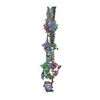
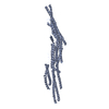
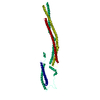
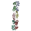
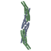
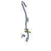
| Title | Crystal structure of the carboxy-terminal region of the bacteriophage T4 proximal long tail fibre protein gp34, R32 native crystal | ||||||
 Components Components | LARGE TAIL FIBER PROTEIN P34 | ||||||
 Keywords Keywords | VIRAL PROTEIN / CAUDOVIRALES / MYOVIRIDAE / TRIPLE BETA-HELIX | ||||||
| Function / homology | : / Long-tail fiber proximal subunit, C-terminal, second / : / Long-tail fiber proximal subunit, C-terminal, trimerization domain / virus tail, fiber / Long-tail fiber proximal subunit Function and homology information Function and homology information | ||||||
| Biological species |  ENTEROBACTERIA PHAGE T4 (virus) ENTEROBACTERIA PHAGE T4 (virus) | ||||||
| Method |  X-RAY DIFFRACTION / SYNCHROTRON / MOLECULAR REPLACEMENT / Resolution: 3 Å X-RAY DIFFRACTION / SYNCHROTRON / MOLECULAR REPLACEMENT / Resolution: 3 Å | ||||||
 Authors Authors | Granell, M. / Alvira, S. / Garcia-Doval, C. / Singh, A.K. / van Raaij, M.J. | ||||||
 Citation Citation | Journal: Viruses / Year: 2017 Title: Crystal Structure of the Carboxy-Terminal Region of the Bacteriophage T4 Proximal Long Tail Fiber Protein Gp34. Authors: Granell, M. / Namura, M. / Alvira, S. / Kanamaru, S. / van Raaij, M.J. #1: Journal: Acta Crystallogr.,Sect.F / Year: 2014 Title: Crystallization of the Carboxy-Terminal Region of the Bacteriophage T4 Proximal Long Tail Fibre Protein Gp34. Authors: Granell, M. / Namura, M. / Alvira, S. / Garcia-Doval, C. / Singh, A.K. / Gutsche, I. / Van Raaij, M.J. / Kanamaru, S. | ||||||
| History |
| ||||||
| Remark 700 | SHEET THE SHEET STRUCTURE OF THIS MOLECULE IS BIFURCATED. IN ORDER TO REPRESENT THIS FEATURE IN ... SHEET THE SHEET STRUCTURE OF THIS MOLECULE IS BIFURCATED. IN ORDER TO REPRESENT THIS FEATURE IN THE SHEET RECORDS BELOW, TWO SHEETS ARE DEFINED. |
- Structure visualization
| Structure viewer | Molecule: MolmilJmol/JSmol |
|---|
- Downloads & links
-Download
| PDBx/mmCIF format | 4uxg.cif.gz | 875.3 KB | Display | PDBx/mmCIF format |
|---|---|---|---|---|
| PDB format | pdb4uxg.ent.gz | 731.8 KB | Display | PDB format |
| PDBx/mmJSON format | 4uxg.json.gz | Tree view | PDBx/mmJSON format | |
| Others |  Other downloads Other downloads |
-Validation report
| Arichive directory | https://data.pdbj.org/pub/pdb/validation_reports/ux/4uxgftp://data.pdbj.org/pub/pdb/validation_reports/ux/4uxg | HTTPS FTP |
|---|
-Related structure data
| Related structure data |  4uxeC  4uxfSC  5nxfC  5nxhC C: citing same article ( S: Starting model for refinement |
|---|---|
| Similar structure data |


-Links
 PDBj
PDBj- Assembly
| Deposited unit | 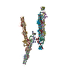
| ||||||||
|---|---|---|---|---|---|---|---|---|---|
| 1 | 
| ||||||||
| 2 | 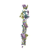
| ||||||||
| 3 | 
| ||||||||
| 4 | 
| ||||||||
| Unit cell |
|
-Components
| #1: Protein | Mass: 44559.973 Da / Num. of mol.: 12 / Fragment: CARBOXY-TERMINAL REGION, RESIDUES 894-1289 Source method: isolated from a genetically manipulated source Source: (gene. exp.) ENTEROBACTERIA PHAGE T4 (virus)Description: GERMAN COLLECTION OF MICROORGANISMS (DSMZ), DSM 4505 Production host:  |
|---|
-Experimental details
-Experiment
| Experiment | Method: X-RAY DIFFRACTION / Number of used crystals: 1 |
|---|
- Sample preparation
| Crystal | Density Matthews: 5 Å3/Da / Density % sol: 76 % / Description: NONE |
|---|---|
| Crystal grow | pH: 8.5 Details: 1.0-1.2 M AMMONIUM SULFATE, 6-16% (V/V) GLYCEROL, 0.1 M TRIS-HCL PH 8.5 |
-Data collection
| Diffraction | Mean temperature: 100 K |
|---|---|
| Diffraction source | Source: SYNCHROTRON / Site: ESRF  / Beamline: ID14-4 / Wavelength: 0.9393 / Beamline: ID14-4 / Wavelength: 0.9393 |
| Detector | Type: ADSC QUANTUM 315r / Detector: CCD / Date: Jun 30, 2012 / Details: TOROIDAL FOCUSING MIRROR |
| Radiation | Monochromator: CHANNEL CUT ESRF MONOCHROMATOR SI(111) / Protocol: SINGLE WAVELENGTH / Monochromatic (M) / Laue (L): M / Scattering type: x-ray |
| Radiation wavelength | Wavelength: 0.9393 Å / Relative weight: 1 |
| Reflection | Resolution: 3→30 Å / Num. obs: 214066 / % possible obs: 99.8 % / Redundancy: 4.6 % / Biso Wilson estimate: 55.6 Å2 / Rmerge(I) obs: 0.13 / Net I/σ(I): 8.4 |
| Reflection shell | Resolution: 3→3.16 Å / Redundancy: 4.7 % / Rmerge(I) obs: 0.36 / Mean I/σ(I) obs: 3.5 / % possible all: 99.8 |
- Processing
| Software |
| ||||||||||||||||||||||||||||||||||||||||||
|---|---|---|---|---|---|---|---|---|---|---|---|---|---|---|---|---|---|---|---|---|---|---|---|---|---|---|---|---|---|---|---|---|---|---|---|---|---|---|---|---|---|---|---|
| Refinement | Method to determine structure: MOLECULAR REPLACEMENT Starting model: PDB ENTRY 4UXF Resolution: 3→29.986 Å / SU ML: 0.38 / σ(F): 1.36 / Phase error: 27.79 / Stereochemistry target values: ML
| ||||||||||||||||||||||||||||||||||||||||||
| Solvent computation | Shrinkage radii: 0.9 Å / VDW probe radii: 1.11 Å / Solvent model: FLAT BULK SOLVENT MODEL | ||||||||||||||||||||||||||||||||||||||||||
| Displacement parameters | Biso mean: 51.4 Å2 | ||||||||||||||||||||||||||||||||||||||||||
| Refinement step | Cycle: LAST / Resolution: 3→29.986 Å
| ||||||||||||||||||||||||||||||||||||||||||
| Refine LS restraints |
| ||||||||||||||||||||||||||||||||||||||||||
| LS refinement shell |
|
