 Movie
Movie Controller

[English] 日本語
 Yorodumi
- EMDB-21845: The V1 region of human V-ATPase in state 1 (focused refinement) -
+ Open data

- Basic information
| Entry | Database: EMDB / ID: EMD-21845 | |||||||||
|---|---|---|---|---|---|---|---|---|---|---|
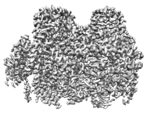
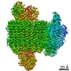
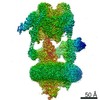
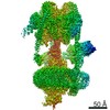
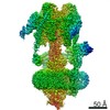
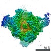
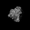
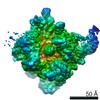
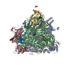
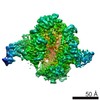
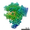
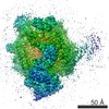
| Title | The V1 region of human V-ATPase in state 1 (focused refinement) | |||||||||
 Map data Map data | The V1 region of human V-ATPase in state 1 (focused refinement) | |||||||||
 Sample Sample |
| |||||||||
 Keywords Keywords | V-ATPase / proton pump / MEMBRANE PROTEIN | |||||||||
| Function / homology |  Function and homology information Function and homology informationproton-transporting two-sector ATPase complex / Ion channel transport / Regulation of MITF-M-dependent genes involved in lysosome biogenesis and autophagy / intracellular pH reduction / symbiont-mediated suppression of host phagosome acidification / ATPase-coupled ion transmembrane transporter activity / Golgi lumen acidification / synaptic vesicle lumen acidification / cellular response to increased oxygen levels / extrinsic component of synaptic vesicle membrane ...proton-transporting two-sector ATPase complex / Ion channel transport / Regulation of MITF-M-dependent genes involved in lysosome biogenesis and autophagy / intracellular pH reduction / symbiont-mediated suppression of host phagosome acidification / ATPase-coupled ion transmembrane transporter activity / Golgi lumen acidification / synaptic vesicle lumen acidification / cellular response to increased oxygen levels / extrinsic component of synaptic vesicle membrane / vacuolar proton-transporting V-type ATPase, V1 domain / Transferrin endocytosis and recycling / lysosomal lumen acidification / clathrin-coated vesicle membrane / endosomal lumen acidification / proton-transporting V-type ATPase complex / Amino acids regulate mTORC1 / protein localization to cilium / vacuolar proton-transporting V-type ATPase complex / ROS and RNS production in phagocytes / vacuolar acidification / proton transmembrane transporter activity / microvillus / proton-transporting ATPase activity, rotational mechanism / cilium assembly / regulation of macroautophagy / H+-transporting two-sector ATPase / specific granule membrane / ATP metabolic process / Insulin receptor recycling / ruffle / secretory granule / proton transmembrane transport / melanosome / synaptic vesicle membrane / ATPase binding / intracellular iron ion homeostasis / endosome / endosome membrane / apical plasma membrane / cilium / Golgi membrane / lysosomal membrane / Neutrophil degranulation / centrosome / ATP hydrolysis activity / extracellular exosome / nucleoplasm / ATP binding / membrane / plasma membrane / cytosol Similarity search - Function | |||||||||
| Biological species |  Homo sapiens (human) / Homo sapiens (human) /  Legionella pneumophila subsp. pneumophila (bacteria) / Legionella pneumophila subsp. pneumophila (strain Philadelphia 1 / ATCC 33152 / DSM 7513) (bacteria) Legionella pneumophila subsp. pneumophila (bacteria) / Legionella pneumophila subsp. pneumophila (strain Philadelphia 1 / ATCC 33152 / DSM 7513) (bacteria) | |||||||||
| Method | single particle reconstruction / cryo EM / Resolution: 2.9 Å | |||||||||
 Authors Authors | Wang L / Wu H | |||||||||
 Citation Citation | Journal: Mol Cell / Year: 2020 Title: Structures of a Complete Human V-ATPase Reveal Mechanisms of Its Assembly. Authors: Longfei Wang / Di Wu / Carol V Robinson / Hao Wu / Tian-Min Fu /   Abstract: Vesicular- or vacuolar-type adenosine triphosphatases (V-ATPases) are ATP-driven proton pumps comprised of a cytoplasmic V complex for ATP hydrolysis and a membrane-embedded V complex for proton ...Vesicular- or vacuolar-type adenosine triphosphatases (V-ATPases) are ATP-driven proton pumps comprised of a cytoplasmic V complex for ATP hydrolysis and a membrane-embedded V complex for proton transfer. They play important roles in acidification of intracellular vesicles, organelles, and the extracellular milieu in eukaryotes. Here, we report cryoelectron microscopy structures of human V-ATPase in three rotational states at up to 2.9-Å resolution. Aided by mass spectrometry, we build all known protein subunits with associated N-linked glycans and identify glycolipids and phospholipids in the V complex. We define ATP6AP1 as a structural hub for V complex assembly because it connects to multiple V subunits and phospholipids in the c-ring. The glycolipids and the glycosylated V subunits form a luminal glycan coat critical for V-ATPase folding, localization, and stability. This study identifies mechanisms of V-ATPase assembly and biogenesis that rely on the integrated roles of ATP6AP1, glycans, and lipids. | |||||||||
| History |
|
- Structure visualization
| Movie |
Movie viewer |
|---|---|
| Structure viewer | EM map: SurfViewMolmilJmol/JSmol |
| Supplemental images |
- Downloads & links
-EMDB archive
| Map data | emd_21845.map.gz | 16.7 MB | EMDB map data format | |
|---|---|---|---|---|
| Header (meta data) | emd-21845-v30.xmlemd-21845.xml | 22.4 KB 22.4 KB | Display Display | EMDB header |
| Images |  emd_21845.png emd_21845.png | 127.3 KB | ||
| Filedesc metadata | emd-21845.cif.gz | 7.4 KB | ||
| Archive directory |  http://ftp.pdbj.org/pub/emdb/structures/EMD-21845ftp://ftp.pdbj.org/pub/emdb/structures/EMD-21845 http://ftp.pdbj.org/pub/emdb/structures/EMD-21845ftp://ftp.pdbj.org/pub/emdb/structures/EMD-21845 | HTTPS FTP |
-Related structure data
| Related structure data |  6wlzMC  6wlwC  6wm2C 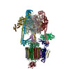 6wm3C  6wm4C M: atomic model generated by this map C: citing same article ( |
|---|---|
| Similar structure data | |
| EM raw data | EMPIAR-11132 (Title: Cryo-EM structures of human V-ATPase / Data size: 8.4 TB Data #1: Unaligned multi frame micrographs of human V-ATPase in complex with SidK [micrographs - multiframe]) |

-Links
| EMDB pages | EMDB (EBI/PDBe) / EMDataResource |
|---|---|
| Related items in Molecule of the Month |


-Map
| File | Download / File: emd_21845.map.gz / Format: CCP4 / Size: 178 MB / Type: IMAGE STORED AS FLOATING POINT NUMBER (4 BYTES) | ||||||||||||||||||||||||||||||||||||||||||||||||||||||||||||||||||||
|---|---|---|---|---|---|---|---|---|---|---|---|---|---|---|---|---|---|---|---|---|---|---|---|---|---|---|---|---|---|---|---|---|---|---|---|---|---|---|---|---|---|---|---|---|---|---|---|---|---|---|---|---|---|---|---|---|---|---|---|---|---|---|---|---|---|---|---|---|---|
| Annotation | The V1 region of human V-ATPase in state 1 (focused refinement) | ||||||||||||||||||||||||||||||||||||||||||||||||||||||||||||||||||||
| Projections & slices | Image control
Images are generated by Spider. | ||||||||||||||||||||||||||||||||||||||||||||||||||||||||||||||||||||
| Voxel size | X=Y=Z: 1.08 Å | ||||||||||||||||||||||||||||||||||||||||||||||||||||||||||||||||||||
| Density |
| ||||||||||||||||||||||||||||||||||||||||||||||||||||||||||||||||||||
| Symmetry | Space group: 1 | ||||||||||||||||||||||||||||||||||||||||||||||||||||||||||||||||||||
| Details | EMDB XML:
CCP4 map header:
| ||||||||||||||||||||||||||||||||||||||||||||||||||||||||||||||||||||
 Z (Sec.)
 Y (Row.)
 X (Col.)


-Supplemental data
- Sample components
+Entire : Human V-ATPase and SidK complex
+Supramolecule #1: Human V-ATPase and SidK complex
+Supramolecule #2: Human V-ATPase
+Supramolecule #3: SidK
+Macromolecule #1: V-type proton ATPase catalytic subunit A
+Macromolecule #2: V-type proton ATPase subunit B, brain isoform
+Macromolecule #3: SidK
+Macromolecule #4: V-type proton ATPase subunit E 1
+Macromolecule #5: V-type proton ATPase subunit G 1
+Macromolecule #6: V-type proton ATPase subunit D
+Macromolecule #7: V-type proton ATPase subunit F
+Macromolecule #8: ADENOSINE-5'-DIPHOSPHATE

-Experimental details
-Structure determination
| Method | cryo EM |
|---|---|
 Processing Processing | single particle reconstruction |
| Aggregation state | particle |
-Sample preparation
| Buffer | pH: 7.4 |
|---|---|
| Vitrification | Cryogen name: ETHANE |
- Electron microscopy
| Microscope | FEI TITAN KRIOS |
|---|---|
| Image recording | Film or detector model: GATAN K3 (6k x 4k) / Average electron dose: 50.1 e/Å2 |
| Electron beam | Acceleration voltage: 300 kV / Electron source:  FIELD EMISSION GUN FIELD EMISSION GUN |
| Electron optics | Illumination mode: SPOT SCAN / Imaging mode: BRIGHT FIELD |
| Experimental equipment |  Model: Titan Krios / Image courtesy: FEI Company |
