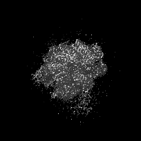
- EMDB-3218: Structure of transcribing mammalian RNA polymerase II (EC1) -
+
Open data
ID or keywords:
Loading...
-
Basic information
Entry
Database: EMDB / ID: EMD-3218
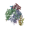
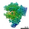
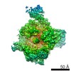
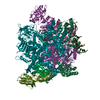
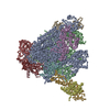
Title
Structure of transcribing mammalian RNA polymerase II (EC1)
Map data
Bovine Pol II elongation complex EC1, B-factor sharpened map
Sample
Sample: bovine Pol II elongation complex
Protein or peptide: DNA-directed RNA polymerase II
Protein or peptide: DNA-directed RNA polymerase II subunit GRINL1A
Other: DNA-RNA synthetic construct
Keywords
transcription / elongation
Function / homology
Function and homology information
Formation of RNA Pol II elongation complex / Formation of the Early Elongation Complex / Transcriptional regulation by small RNAs / FGFR2 alternative splicing / RNA polymerase II transcribes snRNA genes / mRNA Capping / mRNA Splicing - Minor Pathway / RNA Polymerase I Transcription Initiation / RNA Polymerase I Promoter Escape / RNA Polymerase II Promoter Escape ...Formation of RNA Pol II elongation complex / Formation of the Early Elongation Complex / Transcriptional regulation by small RNAs / FGFR2 alternative splicing / RNA polymerase II transcribes snRNA genes / mRNA Capping / mRNA Splicing - Minor Pathway / RNA Polymerase I Transcription Initiation / RNA Polymerase I Promoter Escape / RNA Polymerase II Promoter Escape / RNA Polymerase II Transcription Pre-Initiation And Promoter Opening / RNA Polymerase I Transcription Termination / RNA Polymerase II Transcription Initiation / RNA Polymerase II Transcription Initiation And Promoter Clearance / RNA Polymerase III Transcription Initiation From Type 1 Promoter / RNA Polymerase III Transcription Initiation From Type 2 Promoter / RNA Polymerase III Transcription Initiation From Type 3 Promoter / RNA Pol II CTD phosphorylation and interaction with CE / Estrogen-dependent gene expression / mRNA Polyadenylation / RNA Polymerase II Pre-transcription Events / TP53 Regulates Transcription of DNA Repair Genes / RNA Polymerase II Transcription Elongation / Processing of Capped Intron-Containing Pre-mRNA / B-WICH complex positively regulates rRNA expression / mRNA Splicing - Major Pathway / Formation of TC-NER Pre-Incision Complex / Dual incision in TC-NER / Gap-filling DNA repair synthesis and ligation in TC-NER / DNA/RNA hybrid binding / termination of RNA polymerase II transcription / positive regulation of nuclear-transcribed mRNA poly(A) tail shortening / maintenance of transcriptional fidelity during transcription elongation by RNA polymerase II / positive regulation of translational initiation / nuclear-transcribed mRNA catabolic process / core promoter sequence-specific DNA binding / RNA polymerase I complex / RNA polymerase III complex / transcription elongation by RNA polymerase I / RNA polymerase II, core complex / tRNA transcription by RNA polymerase III / transcription by RNA polymerase I / transcription-coupled nucleotide-excision repair / translation initiation factor binding / transcription initiation at RNA polymerase II promoter / P-body / euchromatin / protein-DNA complex / mRNA transcription by RNA polymerase II / ribonucleoside binding / DNA-directed RNA polymerase / DNA-directed RNA polymerase activity / single-stranded DNA binding / Hydrolases; Acting on ester bonds; Exoribonucleases producing 5'-phosphomonoesters / transcription by RNA polymerase II / nucleic acid binding / chromosome, telomeric region / protein dimerization activity / single-stranded RNA binding / RNA-directed RNA polymerase / nucleotide binding / hydrolase activity / RNA-directed RNA polymerase activity / chromatin binding / nucleolus / magnesium ion binding / DNA binding / zinc ion binding / nucleus / cytosol Similarity search - Function
DNA-directed RNA polymerase II subunit RPB2 / DNA-directed RNA polymerases I, II, and III subunit RPABC3 / DNA-directed RNA polymerase II subunit RPB1 / DNA-directed RNA polymerase II subunit RPB4 / DNA-directed RNA polymerases I, II, and III subunit RPABC1 / DNA-directed RNA polymerase II subunit RPB9 / DNA-directed RNA polymerases I, II, and III subunit RPABC5 / DNA-directed RNA polymerase II subunit RPB11 / DNA-directed RNA polymerases I, II, and III subunit RPABC2 / DNA-directed RNA polymerase II subunit RPB3 ...DNA-directed RNA polymerase II subunit RPB2 / DNA-directed RNA polymerases I, II, and III subunit RPABC3 / DNA-directed RNA polymerase II subunit RPB1 / DNA-directed RNA polymerase II subunit RPB4 / DNA-directed RNA polymerases I, II, and III subunit RPABC1 / DNA-directed RNA polymerase II subunit RPB9 / DNA-directed RNA polymerases I, II, and III subunit RPABC5 / DNA-directed RNA polymerase II subunit RPB11 / DNA-directed RNA polymerases I, II, and III subunit RPABC2 / DNA-directed RNA polymerase II subunit RPB3 / DNA-directed RNA polymerases I, II, and III subunit RPABC4 / DNA-directed RNA polymerase II subunit RPB7 Similarity search - Component
Journal: Nature / Year: 2016 Title: Structure of transcribing mammalian RNA polymerase II. Authors: Carrie Bernecky / Franz Herzog / Wolfgang Baumeister / Jürgen M Plitzko / Patrick Cramer / Abstract: RNA polymerase (Pol) II produces messenger RNA during transcription of protein-coding genes in all eukaryotic cells. The Pol II structure is known at high resolution from X-ray crystallography for ...RNA polymerase (Pol) II produces messenger RNA during transcription of protein-coding genes in all eukaryotic cells. The Pol II structure is known at high resolution from X-ray crystallography for two yeast species. Structural studies of mammalian Pol II, however, remain limited to low-resolution electron microscopy analysis of human Pol II and its complexes with various proteins. Here we report the 3.4 Å resolution cryo-electron microscopy structure of mammalian Pol II in the form of a transcribing complex comprising DNA template and RNA transcript. We use bovine Pol II, which is identical to the human enzyme except for seven amino-acid residues. The obtained atomic model closely resembles its yeast counterpart, but also reveals unknown features. Binding of nucleic acids to the polymerase involves 'induced fit' of the mobile Pol II clamp and active centre region. DNA downstream of the transcription bubble contacts a conserved 'TPSA motif' in the jaw domain of the Pol II subunit RPB5, an interaction that is apparently already established during transcription initiation. Upstream DNA emanates from the active centre cleft at an angle of approximately 105° with respect to downstream DNA. This position of upstream DNA allows for binding of the general transcription elongation factor DSIF (SPT4-SPT5) that we localize over the active centre cleft in a conserved position on the clamp domain of Pol II. Our results define the structure of mammalian Pol II in its functional state, indicate that previous crystallographic analysis of yeast Pol II is relevant for understanding gene transcription in all eukaryotes, and provide a starting point for a mechanistic analysis of human transcription.
History
Deposition
Oct 26, 2015
-
Header (metadata) release
Nov 11, 2015
-
Map release
Jan 20, 2016
-
Update
Feb 3, 2016
-
Current status
Feb 3, 2016
Processing site: PDBe / Status: Released
-
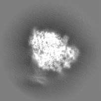
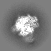
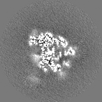
Structure visualization
Movie
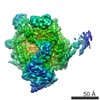
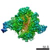
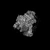
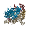
Surface view with section colored by density value
Protein or peptide: DNA-directed RNA polymerase II
Protein or peptide: DNA-directed RNA polymerase II subunit GRINL1A
Other: DNA-RNA synthetic construct
-
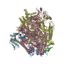
Supramolecule #1000: bovine Pol II elongation complex
Supramolecule
Name: bovine Pol II elongation complex / type: sample / ID: 1000 Details: Recombinant human Gdown1 was present during sample preparation but density was not observed in this map. Number unique components: 3
Molecular weight
Theoretical: 590 KDa
-
Macromolecule #1: DNA-directed RNA polymerase II
Macromolecule
Name: DNA-directed RNA polymerase II / type: protein_or_peptide / ID: 1 / Name.synonym: RNA polymerase II / Number of copies: 1 / Oligomeric state: Monomer / Recombinant expression: No
pH: 7.25 / Details: 150 mM NaCl, 5 mM HEPES, 0.01 mM ZnCl2, 10 mM DTT
Grid
Details: Quantifoil R 3.5/1 holey carbon grids
Vitrification
Cryogen name: ETHANE / Chamber humidity: 100 % / Instrument: FEI VITROBOT MARK IV Method: Four microliters of sample was applied to glow-discharged Quantifoil R 3.5/1 holey carbon grids, which were then blotted for 8.5s and plunge-frozen in liquid ethane.
-
Electron microscopy
Microscope
FEI TITAN KRIOS
Specialist optics
Energy filter - Name: GIF Quantum / Energy filter - Lower energy threshold: 0.0 eV / Energy filter - Upper energy threshold: 20.0 eV
Date
Dec 1, 2014
Image recording
Category: CCD / Film or detector model: GATAN K2 SUMMIT (4k x 4k) / Number real images: 1172 / Average electron dose: 43 e/Å2 Details: Each movie image was collected over 8 s fractionated into 40 frames (0.2 s each).
Electron beam
Acceleration voltage: 300 kV / Electron source: FIELD EMISSION GUN
In the structure databanks used in Yorodumi, some data are registered as the other names, "COVID-19 virus" and "2019-nCoV". Here are the details of the virus and the list of structure data.
Jan 31, 2019. EMDB accession codes are about to change! (news from PDBe EMDB page)
EMDB accession codes are about to change! (news from PDBe EMDB page)
The allocation of 4 digits for EMDB accession codes will soon come to an end. Whilst these codes will remain in use, new EMDB accession codes will include an additional digit and will expand incrementally as the available range of codes is exhausted. The current 4-digit format prefixed with “EMD-” (i.e. EMD-XXXX) will advance to a 5-digit format (i.e. EMD-XXXXX), and so on. It is currently estimated that the 4-digit codes will be depleted around Spring 2019, at which point the 5-digit format will come into force.
The EM Navigator/Yorodumi systems omit the EMD- prefix.
Related info.:Q: What is EMD? / ID/Accession-code notation in Yorodumi/EM Navigator
Yorodumi is a browser for structure data from EMDB, PDB, SASBDB, etc.
This page is also the successor to EM Navigator detail page, and also detail information page/front-end page for Omokage search.
The word "yorodu" (or yorozu) is an old Japanese word meaning "ten thousand". "mi" (miru) is to see.
Related info.:EMDB / PDB / SASBDB / Comparison of 3 databanks / Yorodumi Search / Aug 31, 2016. New EM Navigator & Yorodumi / Yorodumi Papers / Jmol/JSmol / Function and homology information / Changes in new EM Navigator and Yorodumi
 Movie
Movie Controller


 Open data
 Basic information
 Map data
 Sample
 Keywords
 Function and homology information
 Homo sapiens (human) / unidentified (others)
 Authors
 Citation
 Structure visualization
 Downloads & links
 http://ftp.pdbj.org/pub/emdb/structures/EMD-3218
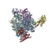


 Z (Sec.)
 Y (Row.)
 X (Col.)


 Sample components
 Processing
 Electron microscopy
 FIELD EMISSION GUN
