National Institutes of Health/National Institute of General Medical Sciences (NIH/NIGMS)
R35GM122510
米国
引用
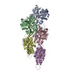
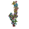
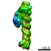
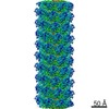
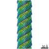
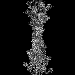
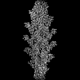
ジャーナル: Bone Res / 年: 2020 タイトル: Osteogenesis imperfecta mutations in plastin 3 lead to impaired calcium regulation of actin bundling. 著者: Christopher L Schwebach / Elena Kudryashova / Weili Zheng / Matthew Orchard / Harper Smith / Lucas A Runyan / Edward H Egelman / Dmitri S Kudryashov / 要旨: Mutations in actin-bundling protein plastin 3 (PLS3) emerged as a cause of congenital osteoporosis, but neither the role of PLS3 in bone development nor the mechanisms underlying PLS3-dependent ...Mutations in actin-bundling protein plastin 3 (PLS3) emerged as a cause of congenital osteoporosis, but neither the role of PLS3 in bone development nor the mechanisms underlying PLS3-dependent osteoporosis are understood. Of the over 20 identified osteoporosis-linked PLS3 mutations, we investigated all five that are expected to produce full-length protein. One of the mutations distorted an actin-binding loop in the second actin-binding domain of PLS3 and abolished F-actin bundling as revealed by cryo-EM reconstruction and protein interaction assays. Surprisingly, the remaining four mutants fully retained F-actin bundling ability. However, they displayed defects in Ca sensitivity: two of the mutants lost the ability to be inhibited by Ca, while the other two became hypersensitive to Ca. Each group of the mutants with similar biochemical properties showed highly characteristic cellular behavior. Wild-type PLS3 was distributed between lamellipodia and focal adhesions. In striking contrast, the Ca-hyposensitive mutants were not found at the leading edge but localized exclusively at focal adhesions/stress fibers, which displayed reinforced morphology. Consistently, the Ca-hypersensitive PLS3 mutants were restricted to lamellipodia, while chelation of Ca caused their redistribution to focal adhesions. Finally, the bundling-deficient mutant failed to co-localize with any F-actin structures in cells despite a preserved F-actin binding through a non-mutation-bearing actin-binding domain. Our findings revealed that severe osteoporosis can be caused by a mutational disruption of the Ca-controlled PLS3's cycling between adhesion complexes and the leading edge. Integration of the structural, biochemical, and cell biology insights enabled us to propose a molecular mechanism of plastin activity regulation by Ca.
 ムービー
ムービー コントローラー


 データを開く
 基本情報
基本情報 マップデータ
 試料
試料 キーワード
キーワード 機能・相同性情報
 Homo sapiens (ヒト)
 データ登録者
 米国, 1件
  引用
引用 構造の表示
構造の表示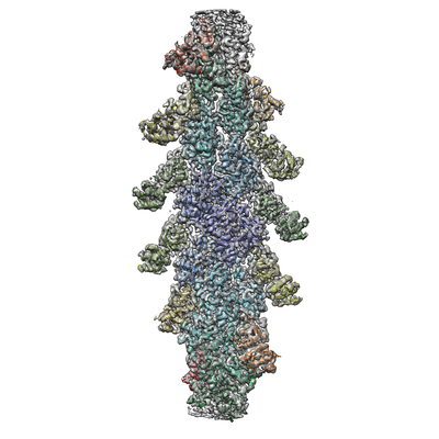
 ダウンロードとリンク
 emd_21155.png
 http://ftp.pdbj.org/pub/emdb/structures/EMD-21155
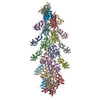


 Z (Sec.)
 Y (Row.)
 X (Col.)


 試料の構成要素

 解析
解析 電子顕微鏡法
 FIELD EMISSION GUN