 Movie
Movie Controller


+ Open data

- Basic information
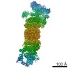
| Entry | Database: EMDB / ID: EMD-6290 | |||||||||
|---|---|---|---|---|---|---|---|---|---|---|
| Title | Negative stain reconstruction of bovine dynactin complex | |||||||||
 Map data Map data | Negative stain reconstruction of bovine dynactin complex | |||||||||
 Sample Sample |
| |||||||||
 Keywords Keywords | dynactin / actin-related proteins / dynein | |||||||||
| Biological species |  | |||||||||
| Method | single particle reconstruction / negative staining / Resolution: 24.0 Å | |||||||||
 Authors Authors | Chowdhury S / Ketcham SA / Schroer TA / Lander GC | |||||||||
 Citation Citation | Journal: Nat Struct Mol Biol / Year: 2015 Title: Structural organization of the dynein-dynactin complex bound to microtubules. Authors: Saikat Chowdhury / Stephanie A Ketcham / Trina A Schroer / Gabriel C Lander /  Abstract: Cytoplasmic dynein associates with dynactin to drive cargo movement on microtubules, but the structure of the dynein-dynactin complex is unknown. Using electron microscopy, we determined the ...Cytoplasmic dynein associates with dynactin to drive cargo movement on microtubules, but the structure of the dynein-dynactin complex is unknown. Using electron microscopy, we determined the organization of native bovine dynein, dynactin and the dynein-dynactin-microtubule quaternary complex. In the microtubule-bound complex, the dynein motor domains are positioned for processive unidirectional movement, and the cargo-binding domains of both dynein and dynactin are accessible. | |||||||||
| History |
|
- Structure visualization
| Movie |
 Movie viewer Movie viewer |
|---|---|
| Structure viewer | EM map: SurfViewMolmilJmol/JSmol |
| Supplemental images |
 UCSF Chimera
- Downloads & links
-EMDB archive
| Map data | emd_6290.map.gz | 576 KB | EMDB map data format | |
|---|---|---|---|---|
| Header (meta data) | emd-6290-v30.xmlemd-6290.xml | 20 KB 20 KB | Display Display | EMDB header |
| Images | 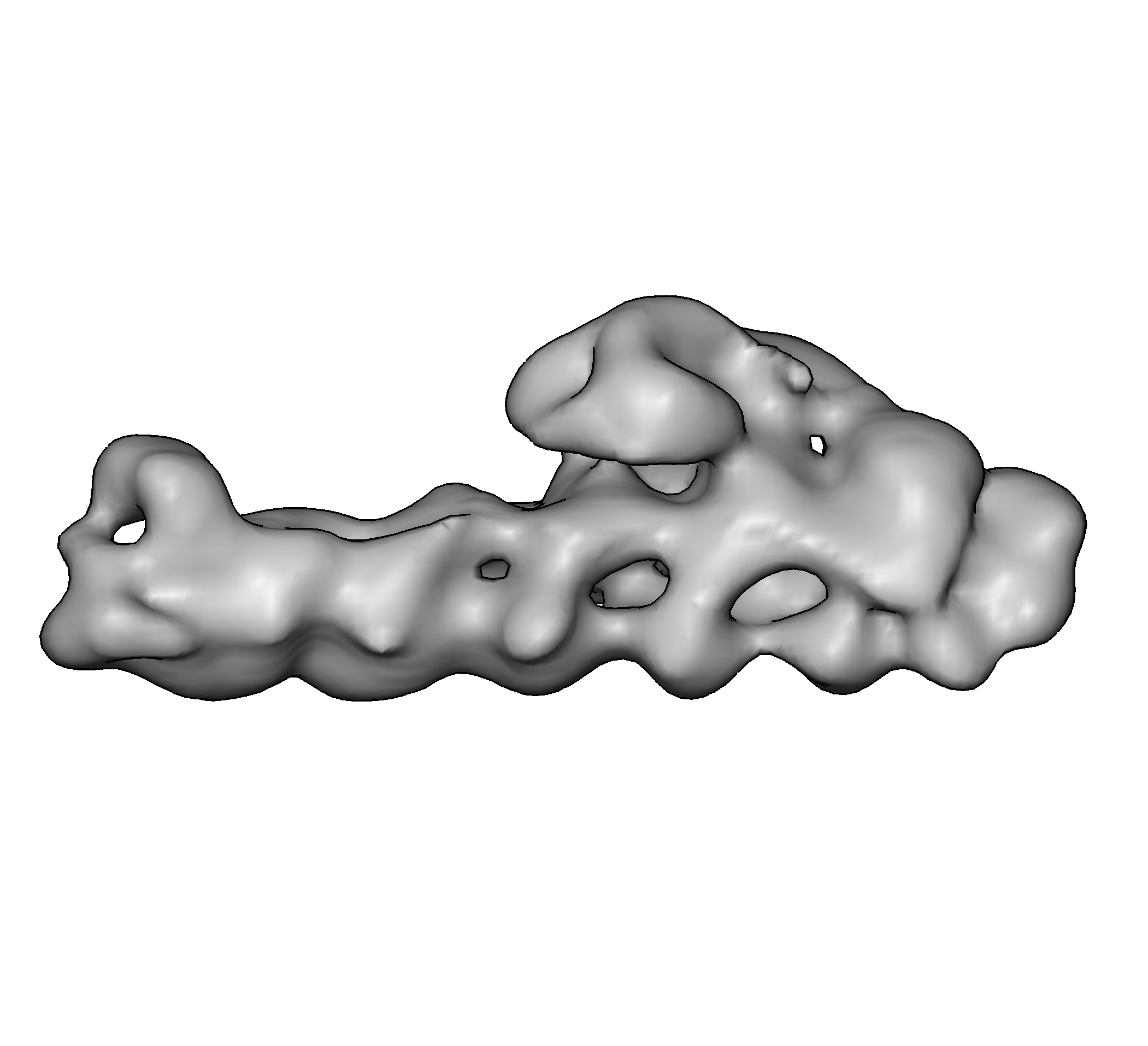 emd_6290.png emd_6290.png | 270.6 KB | ||
| Archive directory |  http://ftp.pdbj.org/pub/emdb/structures/EMD-6290ftp://ftp.pdbj.org/pub/emdb/structures/EMD-6290 http://ftp.pdbj.org/pub/emdb/structures/EMD-6290ftp://ftp.pdbj.org/pub/emdb/structures/EMD-6290 | HTTPS FTP |
-Related structure data
-Links
| EMDB pages | EMDB (EBI/PDBe) / EMDataResource |
|---|
-Map
| File | Download / File: emd_6290.map.gz / Format: CCP4 / Size: 22.5 MB / Type: IMAGE STORED AS FLOATING POINT NUMBER (4 BYTES) | ||||||||||||||||||||||||||||||||||||||||||||||||||||||||||||||||||||
|---|---|---|---|---|---|---|---|---|---|---|---|---|---|---|---|---|---|---|---|---|---|---|---|---|---|---|---|---|---|---|---|---|---|---|---|---|---|---|---|---|---|---|---|---|---|---|---|---|---|---|---|---|---|---|---|---|---|---|---|---|---|---|---|---|---|---|---|---|---|
| Annotation | Negative stain reconstruction of bovine dynactin complex | ||||||||||||||||||||||||||||||||||||||||||||||||||||||||||||||||||||
| Projections & slices | Image control
Images are generated by Spider. | ||||||||||||||||||||||||||||||||||||||||||||||||||||||||||||||||||||
| Voxel size | X=Y=Z: 4.1 Å | ||||||||||||||||||||||||||||||||||||||||||||||||||||||||||||||||||||
| Density |
| ||||||||||||||||||||||||||||||||||||||||||||||||||||||||||||||||||||
| Symmetry | Space group: 1 | ||||||||||||||||||||||||||||||||||||||||||||||||||||||||||||||||||||
| Details | EMDB XML:
CCP4 map header:
| ||||||||||||||||||||||||||||||||||||||||||||||||||||||||||||||||||||
 Z (Sec.)
 Y (Row.)
 X (Col.)


-Supplemental data
- Sample components
+Entire : Bovine dynactin complex
+Supramolecule #1000: Bovine dynactin complex
+Macromolecule #1: p150Glued
+Macromolecule #2: p50
+Macromolecule #3: p24
+Macromolecule #4: Arp1
+Macromolecule #5: Actin
+Macromolecule #6: Arp11
+Macromolecule #7: p62
+Macromolecule #8: p25
+Macromolecule #9: p27
+Macromolecule #10: CapZ alpha
+Macromolecule #11: CapZ beta
-Experimental details
-Structure determination
| Method | negative staining |
|---|---|
 Processing Processing | single particle reconstruction |
| Aggregation state | particle |
-Sample preparation
| Concentration | 0.05 mg/mL |
|---|---|
| Buffer | pH: 7.2 / Details: 35 mM Tris, 5 mM MgSO4, 150 mM KCl, 1mM TCEP |
| Staining | Type: NEGATIVE Details: 4 uL of sample was applied to a freshly plasma-cleaned thin carbon surface that was pre-treated with 0.1% w/v poly-L-lysine hydrobromide. After removing excess protein, negative staining was ...Details: 4 uL of sample was applied to a freshly plasma-cleaned thin carbon surface that was pre-treated with 0.1% w/v poly-L-lysine hydrobromide. After removing excess protein, negative staining was performed with 2% w/v uranyl formate solution. |
| Grid | Details: 400 mesh Cu-Rh Maxtaform grid with a thin continuous carbon film on top |
| Vitrification | Cryogen name: NONE / Instrument: OTHER |
- Electron microscopy
| Microscope | FEI TECNAI SPIRIT |
|---|---|
| Temperature | Min: 294 K / Max: 297 K / Average: 295 K |
| Alignment procedure | Legacy - Astigmatism: Objective astigmatism was corrected using a quadrupole stigmator at 52,000 times magnification. |
| Date | May 1, 2013 |
| Image recording | Category: CCD / Film or detector model: TVIPS TEMCAM-F416 (4k x 4k) / Number real images: 1978 / Average electron dose: 20 e/Å2 Details: Data were collected using Leginon automated image acquisition software. |
| Electron beam | Acceleration voltage: 120 kV / Electron source: LAB6 |
| Electron optics | Calibrated magnification: 52000 / Illumination mode: FLOOD BEAM / Imaging mode: BRIGHT FIELD / Cs: 2.20 mm / Nominal defocus max: 1.5 µm / Nominal defocus min: 0.3 µm / Nominal magnification: 52000 |
| Sample stage | Specimen holder: Room temperature holder / Specimen holder model: SIDE ENTRY, EUCENTRIC |
| Experimental equipment |  Model: Tecnai Spirit / Image courtesy: FEI Company |
-Image processing
| Details | Processing leading up to 3D reconstruction was performed using the Appion package. Particles were selected from micrographs using an automated template-based particle picker. The stack was subjected to five iterations of iterative 2D alignment and classification using multivariate statistical analysis (MSA) and multi-reference alignment (MRA). The clean particle stack was subsequently subjected to 3D refinement by iterative projection matching using EMAN2 and SPARX libraries. |
|---|---|
| CTF correction | Details: Phase flipping of whole micrograph |
| Final reconstruction | Algorithm: OTHER / Resolution.type: BY AUTHOR / Resolution: 24.0 Å / Resolution method: OTHER / Software - Name: EMAN2, SPARX Details: Processing leading up to 3D reconstruction was performed using the Appion package. Particles were selected from micrographs using an automated template-based particle picker. The stack was ...Details: Processing leading up to 3D reconstruction was performed using the Appion package. Particles were selected from micrographs using an automated template-based particle picker. The stack was subjected to five iterations of iterative 2D alignment and classification using multivariate statistical analysis (MSA) and multi-reference alignment (MRA). The clean particle stack was subsequently subjected to 3D refinement by iterative projection matching using EMAN2 and SPARX libraries. Number images used: 46734 |
| Final angle assignment | Details: EMAN2: az 90 degrees, alt 90 degrees, phi 90 degrees |
