 Movie
Movie Controller

[English] 日本語
 Yorodumi
- PDB-1z7y: Crystal Structure of the Arabidopsis thaliana O-Acetylserine Sulf... -
+ Open data

- Basic information
| Entry | Database: PDB / ID: 1z7y | ||||||
|---|---|---|---|---|---|---|---|
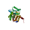
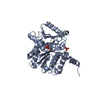
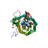
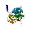
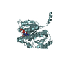
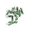
| Title | Crystal Structure of the Arabidopsis thaliana O-Acetylserine Sulfhydrylase K46A mutant | ||||||
 Components Components | Cysteine synthase | ||||||
 Keywords Keywords | TRANSFERASE | ||||||
| Function / homology |  Function and homology information Function and homology informationdouble fertilization forming a zygote and endosperm / pollen tube growth / cysteine synthase / cysteine synthase activity / cysteine biosynthetic process / apoplast / cysteine biosynthetic process from serine / plant-type vacuole / chloroplast stroma / response to cadmium ion ...double fertilization forming a zygote and endosperm / pollen tube growth / cysteine synthase / cysteine synthase activity / cysteine biosynthetic process / apoplast / cysteine biosynthetic process from serine / plant-type vacuole / chloroplast stroma / response to cadmium ion / chloroplast / peroxisome / mRNA binding / nucleus / plasma membrane / cytosol Similarity search - Function | ||||||
| Biological species |  | ||||||
| Method |  X-RAY DIFFRACTION / MOLECULAR REPLACEMENT / Resolution: 2.7 Å X-RAY DIFFRACTION / MOLECULAR REPLACEMENT / Resolution: 2.7 Å | ||||||
 Authors Authors | Bonner, E.R. / Cahoon, R.E. / Knapke, S.M. / Jez, J.M. | ||||||
 Citation Citation | Journal: J.Biol.Chem. / Year: 2005 Title: Molecular Basis of Cysteine Biosynthesis in Plants: STRUCTURAL AND FUNCTIONAL ANALYSIS OF O-ACETYLSERINE SULFHYDRYLASE FROM ARABIDOPSIS THALIANA. Authors: Bonner, E.R. / Cahoon, R.E. / Knapke, S.M. / Jez, J.M. | ||||||
| History |
|
- Structure visualization
| Structure viewer | Molecule: MolmilJmol/JSmol |
|---|
- Downloads & links
-Download
| PDBx/mmCIF format | 1z7y.cif.gz | 74.1 KB | Display | PDBx/mmCIF format |
|---|---|---|---|---|
| PDB format | pdb1z7y.ent.gz | 54.9 KB | Display | PDB format |
| PDBx/mmJSON format | 1z7y.json.gz | Tree view | PDBx/mmJSON format | |
| Others |  Other downloads Other downloads |
-Validation report
| Summary document | 1z7y_validation.pdf.gz | 831.7 KB | Display | wwPDB validaton report |
|---|---|---|---|---|
| Full document | 1z7y_full_validation.pdf.gz | 843.8 KB | Display | |
| Data in XML | 1z7y_validation.xml.gz | 16.2 KB | Display | |
| Data in CIF | 1z7y_validation.cif.gz | 21.7 KB | Display | |
| Arichive directory | https://data.pdbj.org/pub/pdb/validation_reports/z7/1z7yftp://data.pdbj.org/pub/pdb/validation_reports/z7/1z7y | HTTPS FTP |
-Related structure data
| Related structure data |  1z7wC 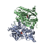 1oasS C: citing same article ( S: Starting model for refinement |
|---|---|
| Similar structure data |

-Links
 PDBj
PDBj
- Assembly
| Deposited unit | 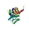
| ||||||||
|---|---|---|---|---|---|---|---|---|---|
| 1 |
| ||||||||
| 2 | 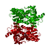
| ||||||||
| Unit cell |
|
-Components
| #1: Protein | Mass: 33781.926 Da / Num. of mol.: 1 / Mutation: K46A Source method: isolated from a genetically manipulated source Source: (gene. exp.)  |
|---|---|
| #2: Chemical | ChemComp-AA5 /   Mass: 378.338 Da / Num. of mol.: 1 / Source method: obtained synthetically / Formula: C13H19N2O7PS Mass: 378.338 Da / Num. of mol.: 1 / Source method: obtained synthetically / Formula: C13H19N2O7PS |
| #3: Water | ChemComp-HOH /  Mass: 18.015 Da / Num. of mol.: 72 / Source method: isolated from a natural source / Formula: H2O Mass: 18.015 Da / Num. of mol.: 72 / Source method: isolated from a natural source / Formula: H2O |
-Experimental details
-Experiment
| Experiment | Method: X-RAY DIFFRACTION / Number of used crystals: 1 |
|---|
- Sample preparation
| Crystal | Density Matthews: 2.03 Å3/Da / Density % sol: 39.48 % |
|---|---|
| Crystal grow | Temperature: 277 K / Method: vapor diffusion, hanging drop / pH: 4.5 Details: sodium formate, pH 4.5, VAPOR DIFFUSION, HANGING DROP, temperature 277K |
-Data collection
| Diffraction | Mean temperature: 100 K |
|---|---|
| Diffraction source | Source: ROTATING ANODE / Type: ENRAF-NONIUS FR591 / Wavelength: 1.5418 |
| Detector | Type: BRUKER SMART 6000 / Detector: CCD / Date: Mar 5, 2005 |
| Radiation | Protocol: SINGLE WAVELENGTH / Monochromatic (M) / Laue (L): M / Scattering type: x-ray |
| Radiation wavelength | Wavelength: 1.5418 Å / Relative weight: 1 |
| Reflection | Resolution: 2.7→60 Å / Num. all: 15955 / Num. obs: 15955 / % possible obs: 98.5 % / Observed criterion σ(I): 0 |
| Reflection shell | Resolution: 2.7→2.8 Å / % possible all: 94.9 |
- Processing
| Software |
| ||||||||||||||||||||
|---|---|---|---|---|---|---|---|---|---|---|---|---|---|---|---|---|---|---|---|---|---|
| Refinement | Method to determine structure: MOLECULAR REPLACEMENT Starting model: PDB Entry 1OAS Resolution: 2.7→15 Å / σ(F): 2 / Stereochemistry target values: Engh & Huber
| ||||||||||||||||||||
| Refinement step | Cycle: LAST / Resolution: 2.7→15 Å
| ||||||||||||||||||||
| Refine LS restraints |
|
