 Movie
Movie Controller


+ Open data

- Basic information
| Entry | Database: PDB / ID: 1w58 | ||||||
|---|---|---|---|---|---|---|---|
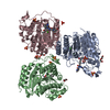
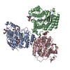
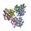
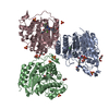
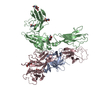
| Title | FtsZ GMPCPP soak I213 (M. jannaschii) | ||||||
 Components Components | CELL DIVISION PROTEIN FTSZ HOMOLOG 1 | ||||||
 Keywords Keywords | CELL DIVISION / CELL DIVISION PROTEIN / CELL-DIVISION PROTEIN / FTSZ / GTP-BINDING / MULTIGENE FAMILY / SEPTATION / TUBULIN | ||||||
| Function / homology |  Function and homology information Function and homology informationFtsZ-dependent cytokinesis / cell division site / protein polymerization / cell division / GTPase activity / GTP binding / cytoplasm Similarity search - Function | ||||||
| Biological species |   METHANOCALDOCOCCUS JANNASCHII (archaea) METHANOCALDOCOCCUS JANNASCHII (archaea) | ||||||
| Method |  X-RAY DIFFRACTION / SYNCHROTRON / MOLECULAR REPLACEMENT / Resolution: 2.5 Å X-RAY DIFFRACTION / SYNCHROTRON / MOLECULAR REPLACEMENT / Resolution: 2.5 Å | ||||||
 Authors Authors | Oliva, M.A. / Cordell, S.C. / Lowe, J. | ||||||
 Citation Citation | Journal: Nat.Struct.Mol.Biol. / Year: 2004 Title: Structural Insights Into Ftsz Protofilament Formation Authors: Oliva, M.A. / Cordell, S.C. / Lowe, J. | ||||||
| History |
|
- Structure visualization
| Structure viewer | Molecule: MolmilJmol/JSmol |
|---|
- Downloads & links
-Download
| PDBx/mmCIF format | 1w58.cif.gz | 81.9 KB | Display | PDBx/mmCIF format |
|---|---|---|---|---|
| PDB format | pdb1w58.ent.gz | 59.9 KB | Display | PDB format |
| PDBx/mmJSON format | 1w58.json.gz | Tree view | PDBx/mmJSON format | |
| Others |  Other downloads Other downloads |
-Validation report
| Arichive directory | https://data.pdbj.org/pub/pdb/validation_reports/w5/1w58ftp://data.pdbj.org/pub/pdb/validation_reports/w5/1w58 | HTTPS FTP |
|---|
-Related structure data
| Related structure data |  1w59C  1w5aC  1w5bC  1w5eC  1w5fC  1fszS S: Starting model for refinement C: citing same article ( |
|---|---|
| Similar structure data |


-Links
 PDBj
PDBj


- Assembly
| Deposited unit | 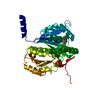
| ||||||||
|---|---|---|---|---|---|---|---|---|---|
| 1 | 
| ||||||||
| Unit cell |
|
-Components
| #1: Protein | Mass: 38970.125 Da / Num. of mol.: 1 Source method: isolated from a genetically manipulated source Source: (gene. exp.) METHANOCALDOCOCCUS JANNASCHII (archaea)Plasmid: PHIS17 / Production host:  |
|---|---|
| #2: Chemical | ChemComp-G2P /   Mass: 521.208 Da / Num. of mol.: 1 / Source method: obtained synthetically / Formula: C11H18N5O13P3 / Comment: GMP-CPP, energy-carrying molecule analogue*YM Mass: 521.208 Da / Num. of mol.: 1 / Source method: obtained synthetically / Formula: C11H18N5O13P3 / Comment: GMP-CPP, energy-carrying molecule analogue*YM |
| #3: Chemical | ChemComp-MG /   Mass: 24.305 Da / Num. of mol.: 1 / Source method: obtained synthetically / Formula: Mg Mass: 24.305 Da / Num. of mol.: 1 / Source method: obtained synthetically / Formula: Mg |
| #4: Water | ChemComp-HOH /  Mass: 18.015 Da / Num. of mol.: 138 / Source method: isolated from a natural source / Formula: H2O Mass: 18.015 Da / Num. of mol.: 138 / Source method: isolated from a natural source / Formula: H2O |
-Experimental details
-Experiment
| Experiment | Method: X-RAY DIFFRACTION / Number of used crystals: 1 |
|---|
- Sample preparation
| Crystal | Density Matthews: 4.48 Å3/Da / Density % sol: 70 % |
|---|
-Data collection
| Diffraction | Mean temperature: 100 K |
|---|---|
| Diffraction source | Source: SYNCHROTRON / Site: SRS  / Beamline: PX14.1 / Wavelength: 0.87 / Beamline: PX14.1 / Wavelength: 0.87 |
| Radiation | Protocol: SINGLE WAVELENGTH / Monochromatic (M) / Laue (L): M / Scattering type: x-ray |
| Radiation wavelength | Wavelength: 0.87 Å / Relative weight: 1 |
| Reflection | Resolution: 2.5→50 Å / Num. obs: 19596 / % possible obs: 98.9 % / Observed criterion σ(I): 0 / Redundancy: 3.2 % / Rmerge(I) obs: 0.06 / Net I/σ(I): 12.8 |
| Reflection shell | Resolution: 2.5→2.65 Å / Redundancy: 2.9 % / Rmerge(I) obs: 0.22 / Mean I/σ(I) obs: 3.2 / % possible all: 99.2 |
- Processing
| Software |
| ||||||||||||||||||||||||||||||||||||||||||||||||||||||||||||
|---|---|---|---|---|---|---|---|---|---|---|---|---|---|---|---|---|---|---|---|---|---|---|---|---|---|---|---|---|---|---|---|---|---|---|---|---|---|---|---|---|---|---|---|---|---|---|---|---|---|---|---|---|---|---|---|---|---|---|---|---|---|
| Refinement | Method to determine structure: MOLECULAR REPLACEMENT Starting model: PDB ENTRY 1FSZ Resolution: 2.5→30 Å / Data cutoff high absF: 10000 / Cross valid method: THROUGHOUT / σ(F): 0 / Stereochemistry target values: MLF
| ||||||||||||||||||||||||||||||||||||||||||||||||||||||||||||
| Solvent computation | Bsol: 66.9844 Å2 / ksol: 0.34496 e/Å3 | ||||||||||||||||||||||||||||||||||||||||||||||||||||||||||||
| Refinement step | Cycle: LAST / Resolution: 2.5→30 Å
| ||||||||||||||||||||||||||||||||||||||||||||||||||||||||||||
| Refine LS restraints |
| ||||||||||||||||||||||||||||||||||||||||||||||||||||||||||||
| LS refinement shell | Resolution: 2.5→2.54 Å / Total num. of bins used: 24 /
| ||||||||||||||||||||||||||||||||||||||||||||||||||||||||||||
| Xplor file |
|
