 Movie
Movie Controller

[English] 日本語
 Yorodumi
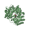
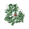
- PDB-2isq: Crystal Structure of O-Acetylserine Sulfhydrylase from Arabidopsi... -
+ Open data

- Basic information
| Entry | Database: PDB / ID: 2isq | ||||||
|---|---|---|---|---|---|---|---|
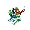
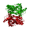
| Title | Crystal Structure of O-Acetylserine Sulfhydrylase from Arabidopsis Thaliana in Complex with C-Terminal Peptide from Arabidopsis Serine Acetyltransferase | ||||||
 Components Components |
| ||||||
 Keywords Keywords | TRANSFERASE / alpha beta structrual domain | ||||||
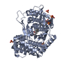
| Function / homology |  Function and homology information Function and homology informationnegative regulation of leaf senescence / double fertilization forming a zygote and endosperm / pollen tube growth / cellular response to sulfate starvation / L-serine O-acetyltransferase activity / serine O-acetyltransferase / cysteine synthase / L-cysteine biosynthetic process / cysteine synthase activity / apoplast ...negative regulation of leaf senescence / double fertilization forming a zygote and endosperm / pollen tube growth / cellular response to sulfate starvation / L-serine O-acetyltransferase activity / serine O-acetyltransferase / cysteine synthase / L-cysteine biosynthetic process / cysteine synthase activity / apoplast / : / plant-type vacuole / chloroplast stroma / response to cadmium ion / chloroplast / peroxisome / mRNA binding / nucleus / plasma membrane / cytosol / cytoplasm Similarity search - Function | ||||||
| Biological species |  | ||||||
| Method |  X-RAY DIFFRACTION / MOLECULAR REPLACEMENT / Resolution: 2.8 Å X-RAY DIFFRACTION / MOLECULAR REPLACEMENT / Resolution: 2.8 Å | ||||||
 Authors Authors | Francois, J.A. / Kumaran, S. / Jez, J.M. | ||||||
 Citation Citation | Journal: Plant Cell / Year: 2006 Title: Structural basis for interaction of o-acetylserine sulfhydrylase and serine acetyltransferase in the Arabidopsis cysteine synthase complex. Authors: Francois, J.A. / Kumaran, S. / Jez, J.M. | ||||||
| History |
|
- Structure visualization
| Structure viewer | Molecule: MolmilJmol/JSmol |
|---|
- Downloads & links
-Download
| PDBx/mmCIF format | 2isq.cif.gz | 77.4 KB | Display | PDBx/mmCIF format |
|---|---|---|---|---|
| PDB format | pdb2isq.ent.gz | 57.2 KB | Display | PDB format |
| PDBx/mmJSON format | 2isq.json.gz | Tree view | PDBx/mmJSON format | |
| Others |  Other downloads Other downloads |
-Validation report
| Arichive directory | https://data.pdbj.org/pub/pdb/validation_reports/is/2isqftp://data.pdbj.org/pub/pdb/validation_reports/is/2isq | HTTPS FTP |
|---|
-Related structure data
| Related structure data | 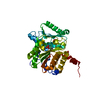 1z7wS S: Starting model for refinement |
|---|---|
| Similar structure data |
-Links
 PDBj
PDBj


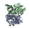
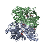
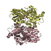
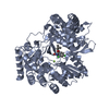
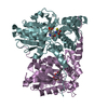
- Assembly
| Deposited unit | 
| ||||||||
|---|---|---|---|---|---|---|---|---|---|
| 1 | 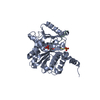
| ||||||||
| Unit cell |
| ||||||||
| Details | Physiologic dimer is generated by crystallographic 2-fold symmetry. |
-Components
| #1: Protein | Mass: 33637.754 Da / Num. of mol.: 1 Source method: isolated from a genetically manipulated source Source: (gene. exp.)  |
|---|---|
| #2: Protein/peptide | Mass: 1012.071 Da / Num. of mol.: 1 / Fragment: SAT peptide Source method: isolated from a genetically manipulated source Source: (gene. exp.) |
| #3: Chemical | ChemComp-SO4 /   Mass: 96.063 Da / Num. of mol.: 1 / Source method: obtained synthetically / Formula: SO4 Mass: 96.063 Da / Num. of mol.: 1 / Source method: obtained synthetically / Formula: SO4 |
| #4: Chemical | ChemComp-PLP /   Mass: 247.142 Da / Num. of mol.: 1 / Source method: obtained synthetically / Formula: C8H10NO6P Mass: 247.142 Da / Num. of mol.: 1 / Source method: obtained synthetically / Formula: C8H10NO6P |
| #5: Water | ChemComp-HOH /  Mass: 18.015 Da / Num. of mol.: 112 / Source method: isolated from a natural source / Formula: H2O Mass: 18.015 Da / Num. of mol.: 112 / Source method: isolated from a natural source / Formula: H2O |
-Experimental details
-Experiment
| Experiment | Method: X-RAY DIFFRACTION / Number of used crystals: 1 |
|---|
- Sample preparation
| Crystal | Density Matthews: 3.94 Å3/Da / Density % sol: 68.76 % |
|---|---|
| Crystal grow | Temperature: 277 K / Method: vapor diffusion, hanging drop / pH: 6.5 Details: 1.5 M ammonium sulfate; 0.1 M PIPES, pH 6.5, VAPOR DIFFUSION, HANGING DROP, temperature 277K |
-Data collection
| Diffraction | Mean temperature: 100 K |
|---|---|
| Diffraction source | Source: ROTATING ANODE / Type: ENRAF-NONIUS FR591 / Wavelength: 1.54 Å |
| Detector | Type: BRUKER SMART 6000 / Detector: CCD / Date: Jun 15, 2006 |
| Radiation | Protocol: SINGLE WAVELENGTH / Monochromatic (M) / Laue (L): M / Scattering type: x-ray |
| Radiation wavelength | Wavelength: 1.54 Å / Relative weight: 1 |
| Reflection | Resolution: 2.8→100 Å / Num. obs: 19828 / % possible obs: 100 % / Observed criterion σ(I): 0 / Redundancy: 10.8 % / Rsym value: 0.145 / Net I/σ(I): 7.36 |
| Reflection shell | Resolution: 2.8→2.9 Å / Redundancy: 10 % / Mean I/σ(I) obs: 2.01 / Num. unique all: 1381 / Rsym value: 0.526 / % possible all: 100 |
- Processing
| Software |
| |||||||||||||||||||||||||
|---|---|---|---|---|---|---|---|---|---|---|---|---|---|---|---|---|---|---|---|---|---|---|---|---|---|---|
| Refinement | Method to determine structure: MOLECULAR REPLACEMENT Starting model: PDB 1Z7W Resolution: 2.8→100 Å / Cross valid method: THROUGHOUT / σ(F): 0 / Stereochemistry target values: Engh & Huber
| |||||||||||||||||||||||||
| Refinement step | Cycle: LAST / Resolution: 2.8→100 Å
| |||||||||||||||||||||||||
| Refine LS restraints |
|
