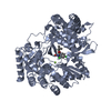
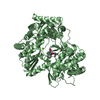
[English] 日本語
Yorodumi
- PDB-3gol: HCV NS5b polymerase in complex with 1,5 benzodiazepine inhibitor ... -

+
Open data


ID or keywords:

Loading...

-
Basic information

Entry
Database: PDB / ID: 3gol
TitleHCV NS5b polymerase in complex with 1,5 benzodiazepine inhibitor (R)-11d
ComponentsRNA-directed RNA polymeraseRNA-dependent RNA polymerase
KeywordsTRANSFERASE / 1 / 5-Benzodiazepine / HCV Polymerase / NS5B / medicinal chemistry / 1b-J4
Function / homology
Function and homology information


hepacivirin / host cell mitochondrial membrane / host cell lipid droplet / symbiont-mediated suppression of host TRAF-mediated signal transduction / transformation of host cell by virus / symbiont-mediated perturbation of host cell cycle G1/S transition checkpoint / symbiont-mediated suppression of host JAK-STAT cascade via inhibition of STAT1 activity / symbiont-mediated suppression of host cytoplasmic pattern recognition receptor signaling pathway via inhibition of MAVS activity / SH3 domain binding / : ...hepacivirin / host cell mitochondrial membrane / host cell lipid droplet / symbiont-mediated suppression of host TRAF-mediated signal transduction / transformation of host cell by virus / symbiont-mediated perturbation of host cell cycle G1/S transition checkpoint / symbiont-mediated suppression of host JAK-STAT cascade via inhibition of STAT1 activity / symbiont-mediated suppression of host cytoplasmic pattern recognition receptor signaling pathway via inhibition of MAVS activity / SH3 domain binding / : / nucleoside-triphosphate phosphatase / protein complex oligomerization / monoatomic ion channel activity / clathrin-dependent endocytosis of virus by host cell / viral nucleocapsid / Hydrolases; Acting on peptide bonds (peptidases); Cysteine endopeptidases / RNA helicase activity / host cell endoplasmic reticulum membrane / host cell perinuclear region of cytoplasm / molecular adaptor activity / RNA helicase / induction by virus of host autophagy / ribonucleoprotein complex / RNA-directed RNA polymerase / viral RNA genome replication / cysteine-type endopeptidase activity / RNA-dependent RNA polymerase activity / serine-type endopeptidase activity / fusion of virus membrane with host endosome membrane / viral envelope / symbiont-mediated suppression of host type I interferon-mediated signaling pathway / host cell nucleus / virion attachment to host cell / host cell plasma membrane / virion membrane / structural molecule activity / ATP hydrolysis activity / proteolysis / RNA binding / zinc ion binding / ATP binding / membrane
Similarity search - Function
Hepatitis C virus core protein, chain A superfamily / Hepatitus C virus, Non-structural 5a protein, C-terminal / Hepatitis C virus NS5A, 1B domain superfamily / Hepatitis C virus non-structural protein NS2, C-terminal domain / Hepatitis C virus non-structural protein NS2, N-terminal domain / Hepatitis C virus non-structural protein NS2 / HCV NS5a protein C-terminal region / Hepatitis C virus, Non-structural protein NS4b / Hepatitis C virus, Core protein, N-terminal / Hepatitis C virus non-structural protein NS4b ...Hepatitis C virus core protein, chain A superfamily / Hepatitus C virus, Non-structural 5a protein, C-terminal / Hepatitis C virus NS5A, 1B domain superfamily / Hepatitis C virus non-structural protein NS2, C-terminal domain / Hepatitis C virus non-structural protein NS2, N-terminal domain / Hepatitis C virus non-structural protein NS2 / HCV NS5a protein C-terminal region / Hepatitis C virus, Non-structural protein NS4b / Hepatitis C virus, Core protein, N-terminal / Hepatitis C virus non-structural protein NS4b / Hepatitis C virus capsid protein / Hepatitis C virus, Non-structural protein NS2 / Hepatitis C virus, Non-structural 5a protein / Hepatitis C virus, Non-structural 5a protein, domain 1a / Hepatitis C virus non-structural 5a, 1B domain / NS5A domain 1a superfamily / Hepatitis C virus non-structural 5a protein membrane anchor / Hepatitis C virus non-structural 5a zinc finger domain / Hepatitis C virus non-structural 5a domain 1b / Hepacivirus nonstructural protein 2 (NS2) protease domain profile. / Hepatitis C virus, Non-structural protein NS4a / Hepatitis C virus non-structural protein NS4a / Hepatitis C virus, Core protein, C-terminal / Hepatitis C virus core protein / Hepatitis C virus, Non-structural protein E2/NS1 / Hepatitis C virus non-structural protein E2/NS1 / Hepatitis C virus, Envelope glycoprotein E1 / Hepatitis C virus envelope glycoprotein E1 / RNA dependent RNA polymerase, hepatitis C virus / Viral RNA dependent RNA polymerase / Hepatitis C virus, NS3 protease, Peptidase S29 / Hepatitis C virus NS3 protease / Hepacivirus/Pegivirus NS3 protease domain profile. / DEAD box, Flavivirus / Flavivirus DEAD domain / Superfamilies 1 and 2 helicase C-terminal domain profile. / Superfamilies 1 and 2 helicase ATP-binding type-1 domain profile. / DEAD-like helicases superfamily / Helicase, C-terminal / Helicase superfamily 1/2, ATP-binding domain / Reverse transcriptase/Diguanylate cyclase domain / RNA-directed RNA polymerase, catalytic domain / RdRp of positive ssRNA viruses catalytic domain profile. / Peptidase S1, PA clan, chymotrypsin-like fold / Peptidase S1, PA clan / DNA/RNA polymerase superfamily / P-loop containing nucleoside triphosphate hydrolase
Similarity search - Domain/homology
Chem-XND / Genome polyprotein
Similarity search - Component
Biological speciesHepatitis C virus isolate
MethodX-RAY DIFFRACTION / SYNCHROTRON / MOLECULAR REPLACEMENT / Resolution: 2.85 Å
AuthorsNyanguile, O. / De Bondt, H.
CitationJournal: Bioorg.Med.Chem.Lett. / Year: 2009
Title: 1,5-Benzodiazepine inhibitors of HCV NS5B polymerase.
Authors: McGowan, D. / Nyanguile, O. / Cummings, M.D. / Vendeville, S. / Vandyck, K. / Van den Broeck, W. / Boutton, C.W. / De Bondt, H. / Quirynen, L. / Amssoms, K. / Bonfanti, J.F. / Last, S. / ...Authors: McGowan, D. / Nyanguile, O. / Cummings, M.D. / Vendeville, S. / Vandyck, K. / Van den Broeck, W. / Boutton, C.W. / De Bondt, H. / Quirynen, L. / Amssoms, K. / Bonfanti, J.F. / Last, S. / Rombauts, K. / Tahri, A. / Hu, L. / Delouvroy, F. / Vermeiren, K. / Vandercruyssen, G. / Van der Helm, L. / Cleiren, E. / Mostmans, W. / Lory, P. / Pille, G. / Van Emelen, K. / Fanning, G. / Pauwels, F. / Lin, T.I. / Simmen, K. / Raboisson, P.
History
DepositionMar 19, 2009Deposition site: RCSB / Processing site: PDBJ
Revision 1.0Jun 16, 2009Provider: repository / Type: Initial release
Revision 1.1Jul 13, 2011Group: Source and taxonomy / Version format compliance
Revision 1.2Nov 1, 2017Group: Refinement description / Category: software
Revision 1.3Mar 20, 2024Group: Data collection / Database references / Derived calculations
Category: chem_comp_atom / chem_comp_bond ...chem_comp_atom / chem_comp_bond / database_2 / struct_conn / struct_ref_seq_dif / struct_site
Item: _database_2.pdbx_DOI / _database_2.pdbx_database_accession ..._database_2.pdbx_DOI / _database_2.pdbx_database_accession / _struct_conn.pdbx_dist_value / _struct_conn.ptnr1_auth_asym_id / _struct_conn.ptnr1_auth_comp_id / _struct_conn.ptnr1_auth_seq_id / _struct_conn.ptnr1_label_asym_id / _struct_conn.ptnr1_label_atom_id / _struct_conn.ptnr1_label_comp_id / _struct_conn.ptnr1_label_seq_id / _struct_conn.ptnr2_auth_asym_id / _struct_conn.ptnr2_auth_comp_id / _struct_conn.ptnr2_auth_seq_id / _struct_conn.ptnr2_label_asym_id / _struct_conn.ptnr2_label_atom_id / _struct_conn.ptnr2_label_comp_id / _struct_ref_seq_dif.details / _struct_site.pdbx_auth_asym_id / _struct_site.pdbx_auth_comp_id / _struct_site.pdbx_auth_seq_id

-
Structure visualization

Structure viewerMolecule:
MolmilJmol/JSmol

Downloads & links

-
Assembly

Deposited unit
A: RNA-directed RNA polymerase
B: RNA-directed RNA polymerase
hetero molecules


Theoretical massNumber of molelcules
Total (without water)130,5746
Polymers129,6352
Non-polymers9394
Water2,018112
1
A: RNA-directed RNA polymerase
hetero molecules


Theoretical massNumber of molelcules
Total (without water)65,2873
Polymers64,8171
Non-polymers4702
Water181
TypeNameSymmetry operationNumber
identity operation1_555x,y,z1
MethodPISA
2
B: RNA-directed RNA polymerase
hetero molecules


Theoretical massNumber of molelcules
Total (without water)65,2873
Polymers64,8171
Non-polymers4702
Water181
TypeNameSymmetry operationNumber
identity operation1_555x,y,z1
MethodPISA
Unit cell
Length a, b, c (Å)106.048, 106.620, 134.679
Angle α, β, γ (deg.)90.00, 90.00, 90.00
Int Tables number19
Space group name H-MP212121

-
Components

#1: Protein RNA-directed RNA polymerase / RNA-dependent RNA polymerase / HCV NS5B RNA POLYMERASE / NS5B / p68


Mass: 64817.262 Da / Num. of mol.: 2 / Fragment: UNP residues 2420-2989
Source method: isolated from a genetically manipulated source
Source: (gene. exp.) Hepatitis C virus isolate / Strain: HC-J4 / Gene: NS5B / Plasmid: pET21b / Production host: Escherichia coli (E. coli) / Strain (production host): BL21(DE3) / References: UniProt: O92972, RNA-directed RNA polymerase
#2: Chemical ChemComp-XND / (11R)-10-acetyl-11-(2,4-dichlorophenyl)-6-hydroxy-3,3-dimethyl-2,3,4,5,10,11-hexahydro-1H-dibenzo[b,e][1,4]diazepin-1-one


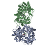
Mass: 445.338 Da / Num. of mol.: 2 / Source method: obtained synthetically / Formula: C23H22Cl2N2O3
#3: Chemical ChemComp-MG / MAGNESIUM ION


Mass: 24.305 Da / Num. of mol.: 2 / Source method: obtained synthetically / Formula: Mg
#4: Water ChemComp-HOH / water / Water


Mass: 18.015 Da / Num. of mol.: 112 / Source method: isolated from a natural source / Formula: H2O

-
Experimental details

-
Experiment

ExperimentMethod: X-RAY DIFFRACTION / Number of used crystals: 1

-
Sample preparation

CrystalDensity Matthews: 2.94 Å3/Da / Density % sol: 58.12 %
Crystal growTemperature: 293 K / Method: vapor diffusion, hanging drop / pH: 6.75
Details: 5-8% PEG 6000, 100mM Mg-Salts, pH 6.75, VAPOR DIFFUSION, HANGING DROP, temperature 293K

-
Data collection

DiffractionMean temperature: 100 K
Diffraction sourceSource: SYNCHROTRON / Site: SLS / Beamline: X10SA / Wavelength: 0.9763 Å
DetectorType: MARMOSAIC 225 mm CCD / Detector: CCD / Date: Jan 1, 2006
RadiationProtocol: SINGLE WAVELENGTH / Monochromatic (M) / Laue (L): M / Scattering type: x-ray
Radiation wavelengthWavelength: 0.9763 Å / Relative weight: 1
ReflectionResolution: 2.85→41.81 Å / Num. all: 36205 / Num. obs: 36205 / % possible obs: 99.6 % / Observed criterion σ(F): 0 / Observed criterion σ(I): 0 / Redundancy: 4.1 % / Rmerge(I) obs: 0.162 / Net I/σ(I): 10.5
Reflection shellResolution: 2.85→2.95 Å / Redundancy: 4.2 % / Rmerge(I) obs: 0.501 / Mean I/σ(I) obs: 3.3 / Num. unique all: 3510 / % possible all: 99.5

-
Processing

Software
NameVersionClassification
XDSdata scaling
REFMAC5.2.0005refinement
XDSdata reduction
XSCALEdata scaling
RefinementMethod to determine structure: MOLECULAR REPLACEMENT / Resolution: 2.85→41.81 Å / Cor.coef. Fo:Fc: 0.928 / Cor.coef. Fo:Fc free: 0.873 / SU B: 27.449 / SU ML: 0.253 / Cross valid method: THROUGHOUT / σ(F): 0 / ESU R Free: 0.369 / Stereochemistry target values: MAXIMUM LIKELIHOOD / Details: HYDROGENS HAVE BEEN ADDED IN THE RIDING POSITIONS
RfactorNum. reflection% reflectionSelection details
Rfree0.2503 1811 5 %RANDOM
Rwork0.1883 ---
all0.19133 34394 --
obs0.19133 34394 99.64 %-
Solvent computationIon probe radii: 0.8 Å / Shrinkage radii: 0.8 Å / VDW probe radii: 1.2 Å / Solvent model: BABINET MODEL WITH MASK
Displacement parametersBiso mean: 33.615 Å2
Baniso -1Baniso -2Baniso -3
1-3.61 Å20 Å20 Å2
2---0.56 Å20 Å2
3----3.05 Å2
Refinement stepCycle: LAST / Resolution: 2.85→41.81 Å
ProteinNucleic acidLigandSolventTotal
Num. atoms8695 0 62 112 8869
Refine LS restraints
Refine-IDTypeDev idealDev ideal targetNumber
X-RAY DIFFRACTIONr_bond_refined_d0.0110.0228950
X-RAY DIFFRACTIONr_bond_other_d0.0010.028178
X-RAY DIFFRACTIONr_angle_refined_deg1.3411.97412158
X-RAY DIFFRACTIONr_angle_other_deg0.785318990
X-RAY DIFFRACTIONr_dihedral_angle_1_deg6.60851111
X-RAY DIFFRACTIONr_dihedral_angle_2_deg36.59122.527364
X-RAY DIFFRACTIONr_dihedral_angle_3_deg19.038151515
X-RAY DIFFRACTIONr_dihedral_angle_4_deg17.8031578
X-RAY DIFFRACTIONr_chiral_restr0.0710.21372
X-RAY DIFFRACTIONr_gen_planes_refined0.0040.029865
X-RAY DIFFRACTIONr_gen_planes_other0.0030.021841
X-RAY DIFFRACTIONr_nbd_refined0.220.22072
X-RAY DIFFRACTIONr_nbd_other0.1820.28439
X-RAY DIFFRACTIONr_nbtor_refined0.1840.24395
X-RAY DIFFRACTIONr_nbtor_other0.0880.25402
X-RAY DIFFRACTIONr_xyhbond_nbd_refined0.1620.2264
X-RAY DIFFRACTIONr_xyhbond_nbd_other0.0210.22
X-RAY DIFFRACTIONr_metal_ion_refined0.0790.22
X-RAY DIFFRACTIONr_metal_ion_other
X-RAY DIFFRACTIONr_symmetry_vdw_refined0.1670.214
X-RAY DIFFRACTIONr_symmetry_vdw_other0.2010.289
X-RAY DIFFRACTIONr_symmetry_hbond_refined0.1450.26
X-RAY DIFFRACTIONr_symmetry_hbond_other
X-RAY DIFFRACTIONr_symmetry_metal_ion_refined
X-RAY DIFFRACTIONr_symmetry_metal_ion_other
X-RAY DIFFRACTIONr_mcbond_it2.00327153
X-RAY DIFFRACTIONr_mcbond_other0.37222235
X-RAY DIFFRACTIONr_mcangle_it2.70839006
X-RAY DIFFRACTIONr_scbond_it4.11143961
X-RAY DIFFRACTIONr_scangle_it6.1663152
X-RAY DIFFRACTIONr_rigid_bond_restr
X-RAY DIFFRACTIONr_sphericity_free
X-RAY DIFFRACTIONr_sphericity_bonded
LS refinement shellResolution: 2.85→2.924 Å / Total num. of bins used: 20
RfactorNum. reflection% reflection
Rfree0.301 131 -
Rwork0.233 2491 -
obs--99.51 %

+
About Yorodumi

-
News

-
Feb 9, 2022. New format data for meta-information of EMDB entries

New format data for meta-information of EMDB entries

  • Version 3 of the EMDB header file is now the official format.
  • The previous official version 1.9 will be removed from the archive.

Related info.:EMDB header

External links:wwPDB to switch to version 3 of the EMDB data model

-
Aug 12, 2020. Covid-19 info

Covid-19 info

URL: https://pdbj.org/emnavi/covid19.php

New page: Covid-19 featured information page in EM Navigator.

Related info.:Covid-19 info / Mar 5, 2020. Novel coronavirus structure data

+
Mar 5, 2020. Novel coronavirus structure data

Novel coronavirus structure data

Related info.:Yorodumi Speices / Aug 12, 2020. Covid-19 info

External links:COVID-19 featured content - PDBj / Molecule of the Month (242):Coronavirus Proteases

+
Jan 31, 2019. EMDB accession codes are about to change! (news from PDBe EMDB page)

EMDB accession codes are about to change! (news from PDBe EMDB page)

  • The allocation of 4 digits for EMDB accession codes will soon come to an end. Whilst these codes will remain in use, new EMDB accession codes will include an additional digit and will expand incrementally as the available range of codes is exhausted. The current 4-digit format prefixed with “EMD-” (i.e. EMD-XXXX) will advance to a 5-digit format (i.e. EMD-XXXXX), and so on. It is currently estimated that the 4-digit codes will be depleted around Spring 2019, at which point the 5-digit format will come into force.
  • The EM Navigator/Yorodumi systems omit the EMD- prefix.

Related info.:Q: What is EMD? / ID/Accession-code notation in Yorodumi/EM Navigator

External links:EMDB Accession Codes are Changing Soon! / Contact to PDBj

+
Jul 12, 2017. Major update of PDB

Major update of PDB

  • wwPDB released updated PDB data conforming to the new PDBx/mmCIF dictionary.
  • This is a major update changing the version number from 4 to 5, and with Remediation, in which all the entries are updated.
  • In this update, many items about electron microscopy experimental information are reorganized (e.g. em_software).
  • Now, EM Navigator and Yorodumi are based on the updated data.

External links:wwPDB Remediation / Enriched Model Files Conforming to OneDep Data Standards Now Available in the PDB FTP Archive

-
Yorodumi

Thousand views of thousand structures

  • Yorodumi is a browser for structure data from EMDB, PDB, SASBDB, etc.
  • This page is also the successor to EM Navigator detail page, and also detail information page/front-end page for Omokage search.
  • The word "yorodu" (or yorozu) is an old Japanese word meaning "ten thousand". "mi" (miru) is to see.

Related info.:EMDB / PDB / SASBDB / Comparison of 3 databanks / Yorodumi Search / Aug 31, 2016. New EM Navigator & Yorodumi / Yorodumi Papers / Jmol/JSmol / Function and homology information / Changes in new EM Navigator and Yorodumi

Read more