 Movie
Movie Controller


+ Open data

- Basic information
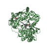
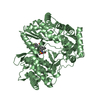
| Entry | Database: PDB / ID: 1nb4 | ||||||
|---|---|---|---|---|---|---|---|
| Title | HC-J4 RNA polymerase apo-form | ||||||
 Components Components | polyprotein | ||||||
 Keywords Keywords | TRANSFERASE / Hepatitis C Virus / replication / RNA polymerase / de-novo priming / function analysis / HCV / NS5B / RDRP | ||||||
| Function / homology |  Function and homology information Function and homology informationhepacivirin / host cell mitochondrial membrane / host cell lipid droplet / symbiont-mediated transformation of host cell / symbiont-mediated suppression of host TRAF-mediated signal transduction / symbiont-mediated perturbation of host cell cycle G1/S transition checkpoint / symbiont-mediated suppression of host JAK-STAT cascade via inhibition of STAT1 activity / symbiont-mediated suppression of host cytoplasmic pattern recognition receptor signaling pathway via inhibition of MAVS activity / SH3 domain binding / nucleoside-triphosphate phosphatase ...hepacivirin / host cell mitochondrial membrane / host cell lipid droplet / symbiont-mediated transformation of host cell / symbiont-mediated suppression of host TRAF-mediated signal transduction / symbiont-mediated perturbation of host cell cycle G1/S transition checkpoint / symbiont-mediated suppression of host JAK-STAT cascade via inhibition of STAT1 activity / symbiont-mediated suppression of host cytoplasmic pattern recognition receptor signaling pathway via inhibition of MAVS activity / SH3 domain binding / nucleoside-triphosphate phosphatase / viral nucleocapsid / channel activity / monoatomic ion transmembrane transport / clathrin-dependent endocytosis of virus by host cell / molecular adaptor activity / Hydrolases; Acting on peptide bonds (peptidases); Cysteine endopeptidases / RNA helicase activity / host cell perinuclear region of cytoplasm / host cell endoplasmic reticulum membrane / RNA helicase / symbiont-mediated suppression of host type I interferon-mediated signaling pathway / ribonucleoprotein complex / serine-type endopeptidase activity / symbiont-mediated activation of host autophagy / RNA-directed RNA polymerase / cysteine-type endopeptidase activity / viral RNA genome replication / RNA-directed RNA polymerase activity / fusion of virus membrane with host endosome membrane / viral envelope / virion attachment to host cell / host cell nucleus / host cell plasma membrane / DNA-templated transcription / virion membrane / structural molecule activity / ATP hydrolysis activity / proteolysis / RNA binding / zinc ion binding / ATP binding Similarity search - Function | ||||||
| Biological species |  Hepatitis C virus Hepatitis C virus | ||||||
| Method |  X-RAY DIFFRACTION / SYNCHROTRON / MOLECULAR REPLACEMENT / Resolution: 2 Å X-RAY DIFFRACTION / SYNCHROTRON / MOLECULAR REPLACEMENT / Resolution: 2 Å | ||||||
 Authors Authors | Jaeger, J. / O'Farrell, D.J. / Trowbridge, R. / Rowlands, D.J. | ||||||
 Citation Citation | Journal: J.Mol.Biol. / Year: 2003 Title: Substrate complexes of hepatitis C virus RNA polymerase (HC-J4): structural evidence for nucleotide import and de-novo initiation. Authors: O'Farrell, D. / Trowbridge, R. / Rowlands, D. / Jager, J. | ||||||
| History |
|
- Structure visualization
| Structure viewer | Molecule: MolmilJmol/JSmol |
|---|
- Downloads & links
-Download
| PDBx/mmCIF format | 1nb4.cif.gz | 243 KB | Display | PDBx/mmCIF format |
|---|---|---|---|---|
| PDB format | pdb1nb4.ent.gz | 194.8 KB | Display | PDB format |
| PDBx/mmJSON format | 1nb4.json.gz | Tree view | PDBx/mmJSON format | |
| Others |  Other downloads Other downloads |
-Validation report
| Arichive directory | https://data.pdbj.org/pub/pdb/validation_reports/nb/1nb4ftp://data.pdbj.org/pub/pdb/validation_reports/nb/1nb4 | HTTPS FTP |
|---|
-Related structure data
| Related structure data | 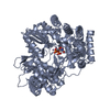 1nb6C  1nb7C  1quvS C: citing same article ( S: Starting model for refinement |
|---|---|
| Similar structure data |


-Links
 PDBj
PDBj

- Assembly
| Deposited unit | 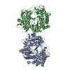
| ||||||||
|---|---|---|---|---|---|---|---|---|---|
| 1 | 
| ||||||||
| 2 | 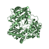
| ||||||||
| Unit cell |
|
-Components
| #1: Protein | Mass: 63456.742 Da / Num. of mol.: 2 / Fragment: RNA dependent RNA polymerase (Residues 2420-2989) Source method: isolated from a genetically manipulated source Source: (gene. exp.) Hepatitis C virus / Genus: Hepacivirus / Plasmid: pET23a / Species (production host): Escherichia coli / Production host:  References: UniProt: Q02828, UniProt: O92972*PLUS, RNA-directed RNA polymerase #2: Water | ChemComp-HOH / |  Mass: 18.015 Da / Num. of mol.: 783 / Source method: isolated from a natural source / Formula: H2O Mass: 18.015 Da / Num. of mol.: 783 / Source method: isolated from a natural source / Formula: H2O |
|---|
-Experimental details
-Experiment
| Experiment | Method: X-RAY DIFFRACTION / Number of used crystals: 1 |
|---|
- Sample preparation
| Crystal | Density Matthews: 2.92 Å3/Da / Density % sol: 56 % | |||||||||||||||||||||||||||||||||||||||||||||||||||||||||||||||
|---|---|---|---|---|---|---|---|---|---|---|---|---|---|---|---|---|---|---|---|---|---|---|---|---|---|---|---|---|---|---|---|---|---|---|---|---|---|---|---|---|---|---|---|---|---|---|---|---|---|---|---|---|---|---|---|---|---|---|---|---|---|---|---|---|
| Crystal grow | Temperature: 291 K / Method: vapor diffusion / pH: 5 Details: PEG 4000, MES, Glycerol, DTT, pH 5.0, VAPOR DIFFUSION, temperature 291K | |||||||||||||||||||||||||||||||||||||||||||||||||||||||||||||||
| Crystal grow | *PLUS Temperature: 18 ℃ / Method: batch method | |||||||||||||||||||||||||||||||||||||||||||||||||||||||||||||||
| Components of the solutions | *PLUS
|
-Data collection
| Diffraction | Mean temperature: 100 K |
|---|---|
| Diffraction source | Source: SYNCHROTRON / Site: SRS  / Beamline: PX14.2 / Wavelength: 1.488 Å / Beamline: PX14.2 / Wavelength: 1.488 Å |
| Detector | Type: ADSC QUANTUM 4 / Detector: CCD / Date: Mar 5, 2001 |
| Radiation | Monochromator: Si 111 CHANNEL / Protocol: SINGLE WAVELENGTH / Monochromatic (M) / Laue (L): M / Scattering type: x-ray |
| Radiation wavelength | Wavelength: 1.488 Å / Relative weight: 1 |
| Reflection | Resolution: 2→20 Å / Num. obs: 99357 / % possible obs: 98.5 % / Redundancy: 5.4 % / Biso Wilson estimate: 15.5 Å2 / Net I/σ(I): 9.1 |
| Reflection shell | Highest resolution: 2 Å / Rsym value: 0.103 |
| Reflection | *PLUS Highest resolution: 2 Å / Lowest resolution: 25 Å / % possible obs: 98.6 % / Redundancy: 5.4 % / Rmerge(I) obs: 0.103 |
- Processing
| Software |
| |||||||||||||||||||||||||
|---|---|---|---|---|---|---|---|---|---|---|---|---|---|---|---|---|---|---|---|---|---|---|---|---|---|---|
| Refinement | Method to determine structure: MOLECULAR REPLACEMENT Starting model: PDB ENTRY 1QUV Resolution: 2→55 Å / Data cutoff high absF: 100000 / Data cutoff high rms absF: 100000 / Cross valid method: THROUGHOUT / σ(F): 2 / Stereochemistry target values: Engh & Huber
| |||||||||||||||||||||||||
| Displacement parameters | Biso mean: 38.5 Å2 | |||||||||||||||||||||||||
| Refine analyze |
| |||||||||||||||||||||||||
| Refinement step | Cycle: LAST / Resolution: 2→55 Å
| |||||||||||||||||||||||||
| Refine LS restraints |
| |||||||||||||||||||||||||
| LS refinement shell | Resolution: 2→2.09 Å / Rfactor Rfree error: 0.012 / Total num. of bins used: 8
| |||||||||||||||||||||||||
| Refinement | *PLUS Lowest resolution: 25 Å / % reflection Rfree: 10 % / Rfactor Rwork: 0.205 | |||||||||||||||||||||||||
| Solvent computation | *PLUS | |||||||||||||||||||||||||
| Displacement parameters | *PLUS | |||||||||||||||||||||||||
| Refine LS restraints | *PLUS
|
