 Movie
Movie Controller

[English] 日本語
 Yorodumi
- PDB-1t03: HIV-1 reverse transcriptase crosslinked to tenofovir terminated t... -
+ Open data

- Basic information
| Entry | Database: PDB / ID: 1t03 | ||||||
|---|---|---|---|---|---|---|---|
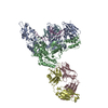
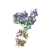
| Title | HIV-1 reverse transcriptase crosslinked to tenofovir terminated template-primer (complex P) | ||||||
 Components Components |
| ||||||
 Keywords Keywords | Transferase/Antibody/DNA / HIV-1 RT / tenofovir / RT-DNA complex / Transferase-Antibody-DNA COMPLEX | ||||||
| Function / homology |  Function and homology information Function and homology informationHIV-1 retropepsin / symbiont-mediated activation of host apoptosis / retroviral ribonuclease H / exoribonuclease H / exoribonuclease H activity / DNA integration / viral genome integration into host DNA / establishment of integrated proviral latency / RNA-directed DNA polymerase / RNA stem-loop binding ...HIV-1 retropepsin / symbiont-mediated activation of host apoptosis / retroviral ribonuclease H / exoribonuclease H / exoribonuclease H activity / DNA integration / viral genome integration into host DNA / establishment of integrated proviral latency / RNA-directed DNA polymerase / RNA stem-loop binding / viral penetration into host nucleus / host multivesicular body / RNA-directed DNA polymerase activity / RNA-DNA hybrid ribonuclease activity / Transferases; Transferring phosphorus-containing groups; Nucleotidyltransferases / host cell / viral nucleocapsid / DNA recombination / DNA-directed DNA polymerase / aspartic-type endopeptidase activity / Hydrolases; Acting on ester bonds / DNA-directed DNA polymerase activity / symbiont-mediated suppression of host gene expression / viral translational frameshifting / symbiont entry into host cell / lipid binding / host cell nucleus / host cell plasma membrane / virion membrane / structural molecule activity / proteolysis / DNA binding / zinc ion binding Similarity search - Function | ||||||
| Biological species |   Human immunodeficiency virus 1 Human immunodeficiency virus 1 | ||||||
| Method |  X-RAY DIFFRACTION / SYNCHROTRON / MOLECULAR REPLACEMENT / Resolution: 3.1 Å X-RAY DIFFRACTION / SYNCHROTRON / MOLECULAR REPLACEMENT / Resolution: 3.1 Å | ||||||
 Authors Authors | Tuske, S. / Sarafianos, S.G. / Ding, J. / Arnold, E. | ||||||
 Citation Citation | Journal: Nat.Struct.Mol.Biol. / Year: 2004 Title: Structure of HIV-1 RT-DNA complexes before and after incorporation of the anti-AIDS drug tenofovir Authors: Tuske, S. / Sarafianos, S.G. / Clark Jr., A.D. / Ding, J. / Naeger, L.K. / White, K.L. / Miller, M.D. / Gibbs, C.S. / Boyer, P.L. / Clark, P. / Wang, G. / Gaffney, B.L. / Jones, R.A. / ...Authors: Tuske, S. / Sarafianos, S.G. / Clark Jr., A.D. / Ding, J. / Naeger, L.K. / White, K.L. / Miller, M.D. / Gibbs, C.S. / Boyer, P.L. / Clark, P. / Wang, G. / Gaffney, B.L. / Jones, R.A. / Jerina, D.M. / Hughes, S.H. / Arnold, E. | ||||||
| History |
| ||||||
| Remark 999 | SEQUENCE The sequence of the FAB heavy and light chains are not available in any of the database ...SEQUENCE The sequence of the FAB heavy and light chains are not available in any of the database sequence. RESIDUE MRG 817 OF THE P CHAIN IS CROSSLINKED TO CHAIN A RESIDUE C258. |
- Structure visualization
| Structure viewer | Molecule: MolmilJmol/JSmol |
|---|
- Downloads & links
-Download
| PDBx/mmCIF format | 1t03.cif.gz | 323.9 KB | Display | PDBx/mmCIF format |
|---|---|---|---|---|
| PDB format | pdb1t03.ent.gz | 254.2 KB | Display | PDB format |
| PDBx/mmJSON format | 1t03.json.gz | Tree view | PDBx/mmJSON format | |
| Others |  Other downloads Other downloads |
-Validation report
| Arichive directory | https://data.pdbj.org/pub/pdb/validation_reports/t0/1t03ftp://data.pdbj.org/pub/pdb/validation_reports/t0/1t03 | HTTPS FTP |
|---|
-Related structure data
| Related structure data | 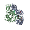 1t05C 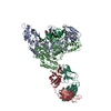 1n5yS S: Starting model for refinement C: citing same article ( |
|---|---|
| Similar structure data |


-Links
 PDBj
PDBj


- Assembly
| Deposited unit | 
| ||||||||
|---|---|---|---|---|---|---|---|---|---|
| 1 |
| ||||||||
| Unit cell |
|
-Components
-Synthetic oligonucleotide ... , 2 types, 2 molecules TP
| #1: DNA chain | Mass: 8367.386 Da / Num. of mol.: 1 / Source method: obtained synthetically |
|---|---|
| #2: DNA chain | Mass: 6430.258 Da / Num. of mol.: 1 / Source method: obtained synthetically |
-Protein , 2 types, 2 molecules AB
| #3: Protein | Mass: 64249.660 Da / Num. of mol.: 1 / Fragment: Reverse transcriptase, p66 subunit / Mutation: Q258C, C280S Source method: isolated from a genetically manipulated source Source: (gene. exp.) Human immunodeficiency virus 1 / Genus: Lentivirus / Gene: POL / Production host:  |
|---|---|
| #4: Protein | Mass: 51095.617 Da / Num. of mol.: 1 / Fragment: Reverse transcriptase, p51 subunit / Mutation: C280S Source method: isolated from a genetically manipulated source Source: (gene. exp.) Human immunodeficiency virus 1 / Genus: Lentivirus / Gene: POL / Production host: |
-Antibody , 2 types, 2 molecules LH
| #5: Antibody | Mass: 23362.650 Da / Num. of mol.: 1 / Fragment: Fab light chain domain / Source method: isolated from a natural source / Source: (natural) |
|---|---|
| #6: Antibody | Mass: 24000.814 Da / Num. of mol.: 1 / Fragment: Fab heavy chain domain / Source method: isolated from a natural source / Source: (natural) |
-Non-polymers , 1 types, 1 molecules 
| #7: Chemical | ChemComp-MG /  Mass: 24.305 Da / Num. of mol.: 1 / Source method: obtained synthetically / Formula: Mg Mass: 24.305 Da / Num. of mol.: 1 / Source method: obtained synthetically / Formula: Mg |
|---|
-Details
| Has protein modification | Y |
|---|
-Experimental details
-Experiment
| Experiment | Method: X-RAY DIFFRACTION / Number of used crystals: 1 |
|---|
- Sample preparation
| Crystal | Density Matthews: 4.98 Å3/Da / Density % sol: 71 % | ||||||||||||||||||||||||||||
|---|---|---|---|---|---|---|---|---|---|---|---|---|---|---|---|---|---|---|---|---|---|---|---|---|---|---|---|---|---|
| Crystal grow | Temperature: 277 K / Method: vapor diffusion, hanging drop / pH: 5.6 Details: 100 mM cacodylate, 33% ammonium sulfate, pH 5.6, VAPOR DIFFUSION, HANGING DROP, temperature 277K | ||||||||||||||||||||||||||||
| Components of the solutions |
|
-Data collection
| Diffraction | Mean temperature: 108 K |
|---|---|
| Diffraction source | Source: SYNCHROTRON / Site: APS  / Beamline: 14-BM-C / Wavelength: 1 Å / Beamline: 14-BM-C / Wavelength: 1 Å |
| Detector | Type: ADSC QUANTUM 4 / Detector: CCD / Date: Feb 21, 2001 |
| Radiation | Monochromator: Bending magnet / Protocol: SINGLE WAVELENGTH / Monochromatic (M) / Laue (L): M / Scattering type: x-ray |
| Radiation wavelength | Wavelength: 1 Å / Relative weight: 1 |
| Reflection | Resolution: 3.1→40 Å / Num. obs: 63979 / % possible obs: 95 % / Observed criterion σ(I): 0 / Biso Wilson estimate: 45 Å2 / Rmerge(I) obs: 0.088 / Net I/σ(I): 12.6 |
| Reflection shell | Resolution: 3.1→3.21 Å / Rmerge(I) obs: 0.388 / Mean I/σ(I) obs: 1.6 / Num. unique all: 4741 / % possible all: 74.5 |
- Processing
| Software |
| |||||||||||||||||||||||||
|---|---|---|---|---|---|---|---|---|---|---|---|---|---|---|---|---|---|---|---|---|---|---|---|---|---|---|
| Refinement | Method to determine structure: MOLECULAR REPLACEMENT Starting model: PDB entry 1N5Y Resolution: 3.1→20 Å / Isotropic thermal model: isotropic / σ(F): 1.1 / Stereochemistry target values: Engh & Huber
| |||||||||||||||||||||||||
| Refine analyze |
| |||||||||||||||||||||||||
| Refinement step | Cycle: LAST / Resolution: 3.1→20 Å
| |||||||||||||||||||||||||
| Refine LS restraints |
| |||||||||||||||||||||||||
| LS refinement shell | Resolution: 3.1→3.12 Å /
|
