 Movie
Movie Controller

[English] 日本語
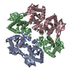
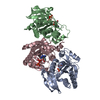
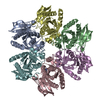
 Yorodumi
- PDB-1pr6: Escherichia coli Purine Nucleoside Phosphorylase Complexed with 9... -
+ Open data

- Basic information
| Entry | Database: PDB / ID: 1pr6 | ||||||
|---|---|---|---|---|---|---|---|
| Title | Escherichia coli Purine Nucleoside Phosphorylase Complexed with 9-beta-D-xylofuranosyladenine and Phosphate/Sulfate | ||||||
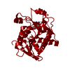
 Components Components | Purine nucleoside phosphorylase DeoD-type | ||||||
 Keywords Keywords | TRANSFERASE / protein-nucleoside complex | ||||||
| Function / homology |  Function and homology information Function and homology informationpurine-nucleoside phosphorylase activity / purine-nucleoside phosphorylase / purine nucleoside catabolic process / cytosol Similarity search - Function | ||||||
| Biological species |  | ||||||
| Method |  X-RAY DIFFRACTION / SYNCHROTRON / MOLECULAR REPLACEMENT / Resolution: 2.1 Å X-RAY DIFFRACTION / SYNCHROTRON / MOLECULAR REPLACEMENT / Resolution: 2.1 Å | ||||||
 Authors Authors | Bennett, E.M. / Li, C. / Allan, P.W. / Parker, W.B. / Ealick, S.E. | ||||||
 Citation Citation | Journal: J.Biol.Chem. / Year: 2003 Title: Structural basis for substrate specificity of Escherichia coli purine nucleoside phosphorylase. Authors: Bennett, E.M. / Li, C. / Allan, P.W. / Parker, W.B. / Ealick, S.E. | ||||||
| History |
|
- Structure visualization
| Structure viewer | Molecule: MolmilJmol/JSmol |
|---|
- Downloads & links
-Download
| PDBx/mmCIF format | 1pr6.cif.gz | 149.8 KB | Display | PDBx/mmCIF format |
|---|---|---|---|---|
| PDB format | pdb1pr6.ent.gz | 118.3 KB | Display | PDB format |
| PDBx/mmJSON format | 1pr6.json.gz | Tree view | PDBx/mmJSON format | |
| Others |  Other downloads Other downloads |
-Validation report
| Arichive directory | https://data.pdbj.org/pub/pdb/validation_reports/pr/1pr6ftp://data.pdbj.org/pub/pdb/validation_reports/pr/1pr6 | HTTPS FTP |
|---|
-Related structure data
| Related structure data | 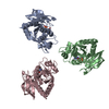 1pk7C 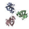 1pk9C 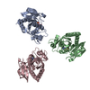 1pkeC 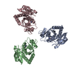 1pr0C  1pr1C  1pr2C  1pr4C  1pr5C  1pw7C 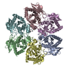 1ecpS S: Starting model for refinement C: citing same article ( |
|---|---|
| Similar structure data |


-Links
 PDBj
PDBj- Assembly
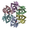
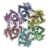
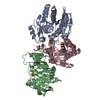
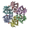
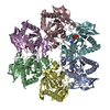
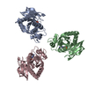
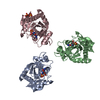
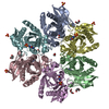
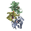
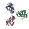
| Deposited unit | 
| ||||||||
|---|---|---|---|---|---|---|---|---|---|
| 1 | 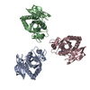
| ||||||||
| Unit cell |
| ||||||||
| Details | The biological hexamer is generated from the trimer in the PDB file by the following operator: x,x-y,1/6-z |
-Components
| #1: Protein | Mass: 25981.947 Da / Num. of mol.: 3 / Source method: isolated from a natural source / Source: (natural) References: UniProt: P0ABP9, purine-nucleoside phosphorylase #2: Chemical |   Mass: 94.971 Da / Num. of mol.: 3 / Source method: obtained synthetically / Formula: PO4 Mass: 94.971 Da / Num. of mol.: 3 / Source method: obtained synthetically / Formula: PO4#3: Chemical | 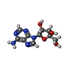  Mass: 267.241 Da / Num. of mol.: 3 / Source method: obtained synthetically / Formula: C10H13N5O4 Mass: 267.241 Da / Num. of mol.: 3 / Source method: obtained synthetically / Formula: C10H13N5O4#4: Water | ChemComp-HOH / |  Mass: 18.015 Da / Num. of mol.: 339 / Source method: isolated from a natural source / Formula: H2O Mass: 18.015 Da / Num. of mol.: 339 / Source method: isolated from a natural source / Formula: H2O |
|---|
-Experimental details
-Experiment
| Experiment | Method: X-RAY DIFFRACTION / Number of used crystals: 1 |
|---|
- Sample preparation
| Crystal | Density Matthews: 3.35 Å3/Da / Density % sol: 63.31 % | ||||||||||||||||||||||||
|---|---|---|---|---|---|---|---|---|---|---|---|---|---|---|---|---|---|---|---|---|---|---|---|---|---|
| Crystal grow | Temperature: 298 K / Method: vapor diffusion, hanging drop / pH: 5.4 Details: ammonium sulfate, citrate, pH 5.4, VAPOR DIFFUSION, HANGING DROP, temperature 298K | ||||||||||||||||||||||||
| Crystal grow | *PLUS Method: vapor diffusion, hanging drop | ||||||||||||||||||||||||
| Components of the solutions | *PLUS
|
-Data collection
| Diffraction | Mean temperature: 100 K |
|---|---|
| Diffraction source | Source: SYNCHROTRON / Site: CHESS  / Beamline: A1 / Wavelength: 0.97 Å / Beamline: A1 / Wavelength: 0.97 Å |
| Detector | Type: ADSC QUANTUM 4 / Detector: CCD / Date: Oct 1, 1996 |
| Radiation | Monochromator: Si 111 / Protocol: SINGLE WAVELENGTH / Monochromatic (M) / Laue (L): M / Scattering type: x-ray |
| Radiation wavelength | Wavelength: 0.97 Å / Relative weight: 1 |
| Reflection | Resolution: 2.1→6 Å / Num. all: 60062 / Num. obs: 53250 / % possible obs: 88.7 % / Observed criterion σ(F): 0 / Observed criterion σ(I): 0 |
| Reflection shell | Resolution: 2.1→2.17 Å / % possible all: 70 |
| Reflection | *PLUS Num. obs: 55490 / % possible obs: 89.2 % / Redundancy: 2.7 % / Rmerge(I) obs: 0.046 |
| Reflection shell | *PLUS Mean I/σ(I) obs: 4.8 |
- Processing
| Software |
| ||||||||||||||||||||
|---|---|---|---|---|---|---|---|---|---|---|---|---|---|---|---|---|---|---|---|---|---|
| Refinement | Method to determine structure: MOLECULAR REPLACEMENT Starting model: PDB entry 1ECP Resolution: 2.1→6 Å / Cross valid method: THROUGHOUT / σ(F): 0 / σ(I): 0 / Stereochemistry target values: Engh & Huber
| ||||||||||||||||||||
| Refinement step | Cycle: LAST / Resolution: 2.1→6 Å
| ||||||||||||||||||||
| Refinement | *PLUS Rfactor Rfree: 0.21 | ||||||||||||||||||||
| Solvent computation | *PLUS | ||||||||||||||||||||
| Displacement parameters | *PLUS | ||||||||||||||||||||
| Refine LS restraints | *PLUS
|
