 Movie
Movie Controller

[English] 日本語
 Yorodumi
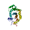
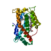
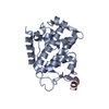
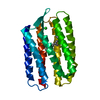
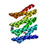
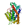
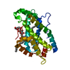
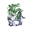
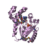
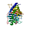
- PDB-1p7m: SOLUTION STRUCTURE AND BASE PERTURBATION STUDIES REVEAL A NOVEL M... -
+ Open data

- Basic information
| Entry | Database: PDB / ID: 1p7m | ||||||
|---|---|---|---|---|---|---|---|
| Title | SOLUTION STRUCTURE AND BASE PERTURBATION STUDIES REVEAL A NOVEL MODE OF ALKYLATED BASE RECOGNITION BY 3-METHYLADENINE DNA GLYCOSYLASE I | ||||||
 Components Components | DNA-3-methyladenine glycosylase I | ||||||
 Keywords Keywords | HYDROLASE / 3-methyladenine TAG complex nmr | ||||||
| Function / homology |  Function and homology information Function and homology informationDNA-3-methyladenine glycosylase I / DNA-3-methyladenine glycosylase activity / DNA alkylation repair / base-excision repair / metal ion binding Similarity search - Function | ||||||
| Biological species |  | ||||||
| Method | SOLUTION NMR / torsion angle dynamics | ||||||
 Authors Authors | Cao, C. / Kwon, K. / Jiang, Y.L. / Drohat, A.C. / Stivers, J.T. | ||||||
 Citation Citation | Journal: J.Biol.Chem. / Year: 2003 Title: Solution structure and base perturbation studies reveal a novel mode of alkylated base recognition by 3-methyladenine DNA glycosylase I Authors: Cao, C. / Kwon, K. / Jiang, Y.L. / Drohat, A.C. / Stivers, J.T. | ||||||
| History |
|
- Structure visualization
| Structure viewer | Molecule: MolmilJmol/JSmol |
|---|
- Downloads & links
-Download
| PDBx/mmCIF format | 1p7m.cif.gz | 1.4 MB | Display | PDBx/mmCIF format |
|---|---|---|---|---|
| PDB format | pdb1p7m.ent.gz | 1.2 MB | Display | PDB format |
| PDBx/mmJSON format | 1p7m.json.gz | Tree view | PDBx/mmJSON format | |
| Others |  Other downloads Other downloads |
-Validation report
| Arichive directory | https://data.pdbj.org/pub/pdb/validation_reports/p7/1p7mftp://data.pdbj.org/pub/pdb/validation_reports/p7/1p7m | HTTPS FTP |
|---|
-Related structure data
| Related structure data | |
|---|---|
| Similar structure data |


-Links
 PDBj
PDBj- Assembly
| Deposited unit | 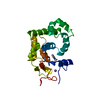
| |||||||||
|---|---|---|---|---|---|---|---|---|---|---|
| 1 |
| |||||||||
| NMR ensembles |
|
-Components
| #1: Protein | Mass: 21138.135 Da / Num. of mol.: 1 Source method: isolated from a genetically manipulated source Source: (gene. exp.) References: UniProt: P05100, DNA-3-methyladenine glycosylase I |
|---|---|
| #2: Chemical | ChemComp-ZN /   Mass: 65.409 Da / Num. of mol.: 1 / Source method: obtained synthetically / Formula: Zn Mass: 65.409 Da / Num. of mol.: 1 / Source method: obtained synthetically / Formula: Zn |
| #3: Chemical | ChemComp-ADK /   Mass: 149.153 Da / Num. of mol.: 1 / Source method: obtained synthetically / Formula: C6H7N5 Mass: 149.153 Da / Num. of mol.: 1 / Source method: obtained synthetically / Formula: C6H7N5 |
-Experimental details
-Experiment
| Experiment | Method: SOLUTION NMR | ||||||||||||||||||||
|---|---|---|---|---|---|---|---|---|---|---|---|---|---|---|---|---|---|---|---|---|---|
| NMR experiment |
| ||||||||||||||||||||
| NMR details | Text: 1H-13C HMQC |
 HSQC
HSQC- Sample preparation
| Details | Contents: 0.5-1mM 13C, 15N, 75% 2H labeled3-methyladenine DNA glycosylase I, 100mM Nacl, 3mM DTT, 0.34mM NaN3, 10mM phosphate buffer, pH6.6 ~8, fold ( or 13C8 labeled) 3-methyladenine Solvent system: H20 |
|---|---|
| Sample conditions | Ionic strength: 10mM phosphate buffer / pH: 6.6 / Pressure: ambient / Temperature: 293 K |
| Crystal grow | *PLUS Method: other / Details: NMR |
-NMR measurement
| Radiation | Protocol: SINGLE WAVELENGTH / Monochromatic (M) / Laue (L): M | ||||||||||||||||||||||||||||||
|---|---|---|---|---|---|---|---|---|---|---|---|---|---|---|---|---|---|---|---|---|---|---|---|---|---|---|---|---|---|---|---|
| Radiation wavelength | Relative weight: 1 | ||||||||||||||||||||||||||||||
| NMR spectrometer |
|
- Processing
| NMR software |
| ||||||||||||||||||||
|---|---|---|---|---|---|---|---|---|---|---|---|---|---|---|---|---|---|---|---|---|---|
| Refinement | Method: torsion angle dynamics / Software ordinal: 1 Details: the structures are based on a total of 2494 restraints, 2140 are NOE-derived distance constraints, 222 dihedral angle restraints, 132 h-bond constraints | ||||||||||||||||||||
| NMR representative | Selection criteria: lowest energy | ||||||||||||||||||||
| NMR ensemble | Conformer selection criteria: structures with acceptable covalent geometry,structures with favorable non-bond energy,structures with the least restraint violations,structures with the lowest energy Conformers calculated total number: 200 / Conformers submitted total number: 25 |
