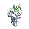
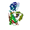
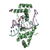
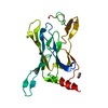
- PDB-1lmz: Solution Structure of 3-Methyladenine DNA Glycosylase I (TAG) -
+
Open data
ID or keywords:
Loading...
-
Basic information
Entry
Database: PDB / ID: 1lmz
Title
Solution Structure of 3-Methyladenine DNA Glycosylase I (TAG)
Components
3-methyladenine DNA glycosylase I (TAG)
Keywords
HYDROLASE / helix-hairpin-helix superfamily / DNA glycosylase / enzyme / TAG / 3-methyladenine / solution structure / NMR spectroscopy
Function / homology
Function and homology information
DNA-3-methyladenine glycosylase I / DNA-3-methyladenine glycosylase activity / DNA alkylation repair / base-excision repair / metal ion binding Similarity search - Function
U-15N-labeled TAG 10 mM phosphate pH 6.6 100 mM NaCl 3 mM DTT 0.34 mM NaN3
93 % water, 7 % deuterium
3
U-13C,15N-labled TAG 10 mM phosphate pH 6.6 100 mM NaCl 3 mM DTT 0.34 mM NaN3
93 % water, 7 % deuterium
Sample conditions
Ionic strength: 100 mM NaCl / pH: 6.6 / Pressure: 1 atm / Temperature: 293 K
Crystal grow
*PLUS
Method: other / Details: NMR
-
NMR measurement
Radiation
Protocol: SINGLE WAVELENGTH / Monochromatic (M) / Laue (L): M / Scattering type: x-ray
Radiation wavelength
Relative weight: 1
NMR spectrometer
Type
Manufacturer
Model
Field strength (MHz)
Spectrometer-ID
Bruker DRX
Bruker
DRX
500
1
Bruker DRX
Bruker
DRX
600
2
Bruker DMX
Bruker
DMX
750
3
Varian UNITYPLUS
Varian
UNITYPLUS
600
4
-
Processing
NMR software
Name
Version
Developer
Classification
CNS
1.1
Brunger, A.T.
structuresolution
NMRPipe
2
Delaglio, F.
processing
Sparky
3
Goddard, T.D., Kneller, D.G.
dataanalysis
XPLOR-NIH
1.1.2
Clore, G.M., Kuszewski, J., Schwieters, C.D., Tjandra, N.
refinement
Refinement
Method: Structures were calculated from an extended strand using CNS 1.1, and refined using XPLOR-NIH 1.1.2 Software ordinal: 1
NMR representative
Selection criteria: lowest energy
NMR ensemble
Conformer selection criteria: structures with acceptable covalent geometry,structures with the least restraint violations,structures with the lowest energy Conformers calculated total number: 100 / Conformers submitted total number: 25
+
About Yorodumi
-
News
-
Feb 9, 2022. New format data for meta-information of EMDB entries
New format data for meta-information of EMDB entries
Version 3 of the EMDB header file is now the official format.
The previous official version 1.9 will be removed from the archive.
In the structure databanks used in Yorodumi, some data are registered as the other names, "COVID-19 virus" and "2019-nCoV". Here are the details of the virus and the list of structure data.
Jan 31, 2019. EMDB accession codes are about to change! (news from PDBe EMDB page)
EMDB accession codes are about to change! (news from PDBe EMDB page)
The allocation of 4 digits for EMDB accession codes will soon come to an end. Whilst these codes will remain in use, new EMDB accession codes will include an additional digit and will expand incrementally as the available range of codes is exhausted. The current 4-digit format prefixed with “EMD-” (i.e. EMD-XXXX) will advance to a 5-digit format (i.e. EMD-XXXXX), and so on. It is currently estimated that the 4-digit codes will be depleted around Spring 2019, at which point the 5-digit format will come into force.
The EM Navigator/Yorodumi systems omit the EMD- prefix.
Related info.:Q: What is EMD? / ID/Accession-code notation in Yorodumi/EM Navigator
Yorodumi is a browser for structure data from EMDB, PDB, SASBDB, etc.
This page is also the successor to EM Navigator detail page, and also detail information page/front-end page for Omokage search.
The word "yorodu" (or yorozu) is an old Japanese word meaning "ten thousand". "mi" (miru) is to see.
Related info.:EMDB / PDB / SASBDB / Comparison of 3 databanks / Yorodumi Search / Aug 31, 2016. New EM Navigator & Yorodumi / Yorodumi Papers / Jmol/JSmol / Function and homology information / Changes in new EM Navigator and Yorodumi
 Movie
Movie Controller


 Open data
 Basic information
 Components
 Keywords
 Function and homology information
 Authors
 Citation
 Structure visualization
 Downloads & links
 Other downloads
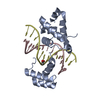


 PDBj
PDBj Assembly
 HNCA
HNCA Sample preparation
 Processing