 Movie
Movie Controller


+ Open data

- Basic information
| Entry | Database: PDB / ID: 4qa8 | ||||||
|---|---|---|---|---|---|---|---|
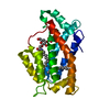
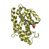
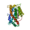
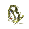
| Title | Crystal structure of LprF from Mycobacterium bovis | ||||||
 Components Components | Putative lipoprotein LprF | ||||||
 Keywords Keywords | LIPID TRANSPORT / lipid transfer / diacylated glycolipid | ||||||
| Function / homology |  Function and homology information Function and homology information | ||||||
| Biological species |  Mycobacterium bovis (bacteria) Mycobacterium bovis (bacteria) | ||||||
| Method |  X-RAY DIFFRACTION / SYNCHROTRON / MOLECULAR REPLACEMENT / Resolution: 1.1 Å X-RAY DIFFRACTION / SYNCHROTRON / MOLECULAR REPLACEMENT / Resolution: 1.1 Å | ||||||
 Authors Authors | Ha, N.C. / Jiao, L. / Kim, J.S. | ||||||
 Citation Citation | Journal: Acta Crystallogr.,Sect.D / Year: 2014 Title: Crystal structure and functional implications of LprF from Mycobacterium tuberculosis and M. bovis Authors: Kim, J.S. / Jiao, L. / Oh, J.I. / Ha, N.C. / Kim, Y.H. | ||||||
| History |
|
- Structure visualization
| Structure viewer | Molecule: MolmilJmol/JSmol |
|---|
- Downloads & links
-Download
| PDBx/mmCIF format | 4qa8.cif.gz | 96 KB | Display | PDBx/mmCIF format |
|---|---|---|---|---|
| PDB format | pdb4qa8.ent.gz | 72.2 KB | Display | PDB format |
| PDBx/mmJSON format | 4qa8.json.gz | Tree view | PDBx/mmJSON format | |
| Others |  Other downloads Other downloads |
-Validation report
| Arichive directory | https://data.pdbj.org/pub/pdb/validation_reports/qa/4qa8ftp://data.pdbj.org/pub/pdb/validation_reports/qa/4qa8 | HTTPS FTP |
|---|
-Related structure data
| Similar structure data |
|---|


-Links
 PDBj
PDBj- Assembly
| Deposited unit | 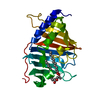
| ||||||||
|---|---|---|---|---|---|---|---|---|---|
| 1 |
| ||||||||
| Unit cell |
|
-Components
| #1: Protein | Mass: 23727.754 Da / Num. of mol.: 1 / Fragment: UNP residues 40-261 Source method: isolated from a genetically manipulated source Source: (gene. exp.) Mycobacterium bovis (bacteria) / Strain: ATCC BAA-935 / AF2122/97 / Gene: lprF, Mb1403 / Production host: |
|---|---|
| #2: Chemical | ChemComp-PJZ / (  Mass: 472.700 Da / Num. of mol.: 1 / Source method: obtained synthetically / Formula: C30H48O4 Mass: 472.700 Da / Num. of mol.: 1 / Source method: obtained synthetically / Formula: C30H48O4 |
| #3: Water | ChemComp-HOH /  Mass: 18.015 Da / Num. of mol.: 156 / Source method: isolated from a natural source / Formula: H2O Mass: 18.015 Da / Num. of mol.: 156 / Source method: isolated from a natural source / Formula: H2O |
-Experimental details
-Experiment
| Experiment | Method: X-RAY DIFFRACTION / Number of used crystals: 1 |
|---|
- Sample preparation
| Crystal | Density Matthews: 2.04 Å3/Da / Density % sol: 39.65 % |
|---|---|
| Crystal grow | Temperature: 295.15 K / Method: vapor diffusion, hanging drop / pH: 7.8 Details: 0.1M Tris-HCl (pH 7.8), 0.2M Magnesium chloride, 28% PEG 4000, VAPOR DIFFUSION, HANGING DROP, temperature 295.15K |
-Data collection
| Diffraction | Mean temperature: 273 K | |||||||||||||||||||||
|---|---|---|---|---|---|---|---|---|---|---|---|---|---|---|---|---|---|---|---|---|---|---|
| Diffraction source | Source: SYNCHROTRON / Site: PAL/PLS  / Beamline: 7A (6B, 6C1) / Wavelength: 0.9789 Å / Beamline: 7A (6B, 6C1) / Wavelength: 0.9789 Å | |||||||||||||||||||||
| Detector | Type: ADSC QUANTUM 270 / Detector: CCD / Date: Jul 23, 2012 | |||||||||||||||||||||
| Radiation | Monochromator: double mirrors / Protocol: SINGLE WAVELENGTH / Monochromatic (M) / Laue (L): M / Scattering type: x-ray | |||||||||||||||||||||
| Radiation wavelength | Wavelength: 0.9789 Å / Relative weight: 1 | |||||||||||||||||||||
| Reflection | Resolution: 1.1→20 Å / Num. all: 68866 / Num. obs: 68768 / % possible obs: 90.4 % / Observed criterion σ(F): 0 / Observed criterion σ(I): 0 | |||||||||||||||||||||
| Reflection shell |
|
- Processing
| Software |
| ||||||||||||||||||||||||||||||||||||||||||||||||||||||||||||||||||||||||||||||||||||||||||||||||||||||||||||||||||||||||||||||||||||||||||||||||||||||||||||
|---|---|---|---|---|---|---|---|---|---|---|---|---|---|---|---|---|---|---|---|---|---|---|---|---|---|---|---|---|---|---|---|---|---|---|---|---|---|---|---|---|---|---|---|---|---|---|---|---|---|---|---|---|---|---|---|---|---|---|---|---|---|---|---|---|---|---|---|---|---|---|---|---|---|---|---|---|---|---|---|---|---|---|---|---|---|---|---|---|---|---|---|---|---|---|---|---|---|---|---|---|---|---|---|---|---|---|---|---|---|---|---|---|---|---|---|---|---|---|---|---|---|---|---|---|---|---|---|---|---|---|---|---|---|---|---|---|---|---|---|---|---|---|---|---|---|---|---|---|---|---|---|---|---|---|---|---|---|
| Refinement | Method to determine structure: MOLECULAR REPLACEMENT / Resolution: 1.1→18.563 Å / SU ML: 0.08 / σ(F): 2.25 / Phase error: 16.82 / Stereochemistry target values: ML
| ||||||||||||||||||||||||||||||||||||||||||||||||||||||||||||||||||||||||||||||||||||||||||||||||||||||||||||||||||||||||||||||||||||||||||||||||||||||||||||
| Solvent computation | Shrinkage radii: 0.9 Å / VDW probe radii: 1.11 Å / Solvent model: FLAT BULK SOLVENT MODEL | ||||||||||||||||||||||||||||||||||||||||||||||||||||||||||||||||||||||||||||||||||||||||||||||||||||||||||||||||||||||||||||||||||||||||||||||||||||||||||||
| Refinement step | Cycle: LAST / Resolution: 1.1→18.563 Å
| ||||||||||||||||||||||||||||||||||||||||||||||||||||||||||||||||||||||||||||||||||||||||||||||||||||||||||||||||||||||||||||||||||||||||||||||||||||||||||||
| Refine LS restraints |
| ||||||||||||||||||||||||||||||||||||||||||||||||||||||||||||||||||||||||||||||||||||||||||||||||||||||||||||||||||||||||||||||||||||||||||||||||||||||||||||
| LS refinement shell | Refine-ID: X-RAY DIFFRACTION / Total num. of bins used: 25
|
