 Movie
Movie Controller


+ Open data

- Basic information
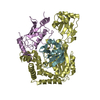
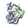
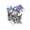
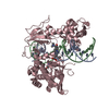
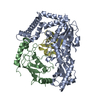
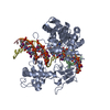
| Entry | Database: PDB / ID: 1nh3 | ||||||
|---|---|---|---|---|---|---|---|
| Title | Human Topoisomerase I Ara-C Complex | ||||||
 Components Components |
| ||||||
 Keywords Keywords | ISOMERASE/DNA / Ara-C / protein-DNA complex / DNA damage / isomerase / ISOMERASE-DNA COMPLEX | ||||||
| Function / homology |  Function and homology information Function and homology informationDNA topoisomerase / DNA topoisomerase type I (single strand cut, ATP-independent) activity / dense fibrillar component / cellular response to luteinizing hormone stimulus / embryonic cleavage / programmed cell death / DNA binding, bending / response to temperature stimulus / supercoiled DNA binding / DNA topological change ...DNA topoisomerase / DNA topoisomerase type I (single strand cut, ATP-independent) activity / dense fibrillar component / cellular response to luteinizing hormone stimulus / embryonic cleavage / programmed cell death / DNA binding, bending / response to temperature stimulus / supercoiled DNA binding / DNA topological change / rRNA transcription / SUMOylation of DNA replication proteins / animal organ regeneration / response to cAMP / male germ cell nucleus / response to gamma radiation / circadian regulation of gene expression / molecular condensate scaffold activity / chromosome segregation / P-body / circadian rhythm / protein-DNA complex / chromatin DNA binding / fibrillar center / single-stranded DNA binding / chromosome / double-stranded DNA binding / perikaryon / DNA replication / RNA polymerase II cis-regulatory region sequence-specific DNA binding / chromatin remodeling / response to xenobiotic stimulus / protein domain specific binding / protein serine/threonine kinase activity / chromatin binding / protein-containing complex binding / nucleolus / DNA binding / RNA binding / nucleoplasm / ATP binding / nucleus Similarity search - Function | ||||||
| Biological species |  Homo sapiens (human) Homo sapiens (human) | ||||||
| Method |  X-RAY DIFFRACTION / SYNCHROTRON / MOLECULAR REPLACEMENT / Resolution: 3.1 Å X-RAY DIFFRACTION / SYNCHROTRON / MOLECULAR REPLACEMENT / Resolution: 3.1 Å | ||||||
 Authors Authors | Chrencik, J.E. / Burgin, A.B. / Pommier, Y. / Stewart, L. / Redinbo, M.R. | ||||||
 Citation Citation | Journal: J.Biol.Chem. / Year: 2003 Title: Structural Impact of the Leukemia Drug 1-beta-D-Arabinofuranosylcytosine (Ara-C) on the Covalent Human Topoisomerase I-DNA Complex Authors: Chrencik, J.E. / Burgin, A.B. / Pommier, Y. / Stewart, L. / Redinbo, M.R. | ||||||
| History |
| ||||||
| Remark 999 | SEQUENCE The DNA (duplex oligo) was added to the protein during crystallization. At this time, the ...SEQUENCE The DNA (duplex oligo) was added to the protein during crystallization. At this time, the protein initiates a transesterification reaction in which one strand of DNA is broken into chains B and C, and the protein chain A is covalently linked to the DNA through residue 723. |
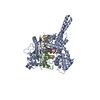
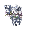
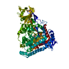
- Structure visualization
| Structure viewer | Molecule: MolmilJmol/JSmol |
|---|
- Downloads & links
-Download
| PDBx/mmCIF format | 1nh3.cif.gz | 137.3 KB | Display | PDBx/mmCIF format |
|---|---|---|---|---|
| PDB format | pdb1nh3.ent.gz | 97.7 KB | Display | PDB format |
| PDBx/mmJSON format | 1nh3.json.gz | Tree view | PDBx/mmJSON format | |
| Others |  Other downloads Other downloads |
-Validation report
| Arichive directory | https://data.pdbj.org/pub/pdb/validation_reports/nh/1nh3ftp://data.pdbj.org/pub/pdb/validation_reports/nh/1nh3 | HTTPS FTP |
|---|
-Related structure data
| Related structure data | 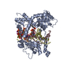 1a31S S: Starting model for refinement |
|---|---|
| Similar structure data |


-Links
 PDBj
PDBj


- Assembly
| Deposited unit | 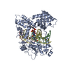
| ||||||||
|---|---|---|---|---|---|---|---|---|---|
| 1 |
| ||||||||
| Unit cell |
|
-Components
| #1: DNA chain | Mass: 3017.004 Da / Num. of mol.: 1 / Source method: obtained synthetically |
|---|---|
| #2: DNA chain | Mass: 3564.368 Da / Num. of mol.: 1 / Source method: obtained synthetically |
| #3: DNA/RNA hybrid | Mass: 6580.128 Da / Num. of mol.: 1 / Source method: obtained synthetically |
| #4: Protein | Mass: 66733.867 Da / Num. of mol.: 1 / Fragment: Core Subdomain, C-Terminal Domain Source method: isolated from a genetically manipulated source Source: (gene. exp.) Homo sapiens (human) / Gene: TOP1 / Cell line (production host): SF9 / Production host:   Spodoptera frugiperda (fall armyworm) / References: UniProt: P11387, EC: 5.99.1.2 Spodoptera frugiperda (fall armyworm) / References: UniProt: P11387, EC: 5.99.1.2 |
| #5: Water | ChemComp-HOH /  Mass: 18.015 Da / Num. of mol.: 33 / Source method: isolated from a natural source / Formula: H2O Mass: 18.015 Da / Num. of mol.: 33 / Source method: isolated from a natural source / Formula: H2O |
| Has protein modification | Y |
-Experimental details
-Experiment
| Experiment | Method: X-RAY DIFFRACTION / Number of used crystals: 1 |
|---|
- Sample preparation
| Crystal | Density Matthews: 3.56 Å3/Da / Density % sol: 65.41 % | ||||||||||||||||||||||||||||||||||||||||||||||||||||||||||||||||||||||
|---|---|---|---|---|---|---|---|---|---|---|---|---|---|---|---|---|---|---|---|---|---|---|---|---|---|---|---|---|---|---|---|---|---|---|---|---|---|---|---|---|---|---|---|---|---|---|---|---|---|---|---|---|---|---|---|---|---|---|---|---|---|---|---|---|---|---|---|---|---|---|---|
| Crystal grow | Temperature: 276 K / Method: vapor diffusion, sitting drop / pH: 7.7 Details: Tris, Magnesium Chloride, PEG 400, DTT, pH 7.7, VAPOR DIFFUSION, SITTING DROP, temperature 276K | ||||||||||||||||||||||||||||||||||||||||||||||||||||||||||||||||||||||
| Crystal grow | *PLUS Temperature: 22 ℃ / Details: Redinbo, M.R., (1998) Science, 279, 1504. | ||||||||||||||||||||||||||||||||||||||||||||||||||||||||||||||||||||||
| Components of the solutions | *PLUS
|
-Data collection
| Diffraction | Mean temperature: 100 K |
|---|---|
| Diffraction source | Source: SYNCHROTRON / Site: NSLS  / Beamline: X12B / Wavelength: 1.1 Å / Beamline: X12B / Wavelength: 1.1 Å |
| Detector | Type: MARRESEARCH / Detector: CCD / Date: Feb 18, 2001 |
| Radiation | Monochromator: SAGITALLY FOCUSED Si(111) / Protocol: SINGLE WAVELENGTH / Monochromatic (M) / Laue (L): M / Scattering type: x-ray |
| Radiation wavelength | Wavelength: 1.1 Å / Relative weight: 1 |
| Reflection | Resolution: 3.1→50 Å / Num. all: 13878 / Num. obs: 13878 / % possible obs: 69.3 % / Observed criterion σ(F): 0 / Observed criterion σ(I): 0 |
| Reflection shell | Resolution: 3.1→3.21 Å / % possible all: 54.7 |
| Reflection | *PLUS Lowest resolution: 100 Å / Redundancy: 12.2 % / Num. measured all: 169504 / Rmerge(I) obs: 0.135 |
| Reflection shell | *PLUS Lowest resolution: 3.2 Å / % possible obs: 54.7 % / Rmerge(I) obs: 0.415 / Mean I/σ(I) obs: 1.4 |
- Processing
| Software |
| |||||||||||||||||||||||||
|---|---|---|---|---|---|---|---|---|---|---|---|---|---|---|---|---|---|---|---|---|---|---|---|---|---|---|
| Refinement | Method to determine structure: MOLECULAR REPLACEMENT Starting model: PDB entry 1A31 Resolution: 3.1→44.29 Å / Cross valid method: THROUGHOUT / σ(F): 0 / Stereochemistry target values: Engh & Huber
| |||||||||||||||||||||||||
| Displacement parameters |
| |||||||||||||||||||||||||
| Refine analyze | Luzzati coordinate error obs: 0.48 Å / Luzzati d res low obs: 5 Å / Luzzati sigma a obs: 0.71 Å | |||||||||||||||||||||||||
| Refinement step | Cycle: LAST / Resolution: 3.1→44.29 Å
| |||||||||||||||||||||||||
| Refine LS restraints |
| |||||||||||||||||||||||||
| Refinement | *PLUS Highest resolution: 3.1 Å / Lowest resolution: 100 Å / % reflection Rfree: 10 % / Rfactor Rfree: 0.243 / Rfactor Rwork: 0.309 | |||||||||||||||||||||||||
| Solvent computation | *PLUS | |||||||||||||||||||||||||
| Displacement parameters | *PLUS | |||||||||||||||||||||||||
| Refine LS restraints | *PLUS
|
