 Movie
Movie Controller

[English] 日本語
 Yorodumi
- PDB-1mae: The Active Site Structure of Methylamine Dehydrogenase: Hydrazine... -
+ Open data

- Basic information
| Entry | Database: PDB / ID: 1mae | ||||||
|---|---|---|---|---|---|---|---|
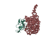
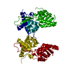
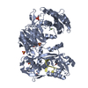
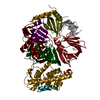
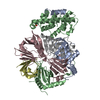
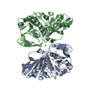
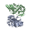
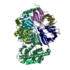
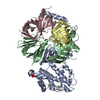
| Title | The Active Site Structure of Methylamine Dehydrogenase: Hydrazines Identify C6 as the Reactive Site of the Tryptophan Derived Quinone Cofactor | ||||||
 Components Components |
| ||||||
 Keywords Keywords | OXIDOREDUCTASE(CHNH2(D)-DEAMINATING) | ||||||
| Function / homology |  Function and homology information Function and homology informationmethylamine dehydrogenase (amicyanin) / methylamine dehydrogenase (amicyanin) activity / methylamine metabolic process / aliphatic amine dehydrogenase activity / amine metabolic process / outer membrane-bounded periplasmic space / periplasmic space Similarity search - Function | ||||||
| Biological species |  Paracoccus versutus (bacteria) Paracoccus versutus (bacteria) | ||||||
| Method |  X-RAY DIFFRACTION / Resolution: 2.8 Å X-RAY DIFFRACTION / Resolution: 2.8 Å | ||||||
 Authors Authors | Huizinga, E.G. / Vellieux, F.M.D. / Hol, W.G.J. | ||||||
 Citation Citation | Journal: Biochemistry / Year: 1992 Title: Active site structure of methylamine dehydrogenase: hydrazines identify C6 as the reactive site of the tryptophan-derived quinone cofactor. Authors: Huizinga, E.G. / van Zanten, B.A. / Duine, J.A. / Jongejan, J.A. / Huitema, F. / Wilson, K.S. / Hol, W.G. #1: Journal: FEBS Lett. / Year: 1991Title: Crystallographic Investigations of the Tryptophan-Derived Cofactor in the Quinoprotein Methylamine Dehydrogenase Authors: Chen, L. / Mathews, F.S. / Davidson, V.L. / Huizinga, E.G. / Vellieux, F.M.D. / Duine, J.A. / Hol, W.G.J. #2: Journal: Embo J. / Year: 1989Title: Structure of Quinoprotein Methylamine Dehydrogenase at 2.25 Angstroms Resolution Authors: Vellieux, F.M.D. / Huitema, F. / Groendijk, H. / Kalk, K.H. / Frank, J. / Jongejan, J.A. / Duine, J.A. / Petratos, K. / Drenth, J. / Hol, W.G.J. #3: Journal: Eur.J.Biochem. / Year: 1986Title: Purification, Crystallization and Preliminary X-Ray Investigation of Quinoprotein Methylamine Dehydrogenase from Thiobacillus Versutus Authors: Vellieux, F.M.D. / Frank, J. / Swarte, M.B.A. / Groendijk, H. / Duine, J.A. / Drenth, J. / Hol, W.G.J. | ||||||
| History |
|
- Structure visualization
| Structure viewer | Molecule: MolmilJmol/JSmol |
|---|
- Downloads & links
-Download
| PDBx/mmCIF format | 1mae.cif.gz | 53.3 KB | Display | PDBx/mmCIF format |
|---|---|---|---|---|
| PDB format | pdb1mae.ent.gz | 35.3 KB | Display | PDB format |
| PDBx/mmJSON format | 1mae.json.gz | Tree view | PDBx/mmJSON format | |
| Others |  Other downloads Other downloads |
-Validation report
| Arichive directory | https://data.pdbj.org/pub/pdb/validation_reports/ma/1maeftp://data.pdbj.org/pub/pdb/validation_reports/ma/1mae | HTTPS FTP |
|---|
-Related structure data


-Links
 PDBj
PDBj- Assembly
| Deposited unit | 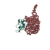
| ||||||||
|---|---|---|---|---|---|---|---|---|---|
| 1 | 
| ||||||||
| Unit cell |
| ||||||||
| Details | THE MOLECULE IS NORMALLY A TETRAMER. THE CRYSTALLOGRAPHIC ASYMMETRIC UNIT CONSISTS OF ONE-HALF OF THE TETRAMER, NAMELY ONE LIGHT CHAIN *L* AND ONE HEAVY CHAIN *H*. TO GENERATE THE FULL MOLECULE, THE FOLLOWING CRYSTALLOGRAPHIC TWO-FOLD OPERATION MUST BE APPLIED TO THE LIGHT AND HEAVY CHAINS PRESENTED IN THIS ENTRY -0.5 0.866025 0.0 0.0 0.866025 0.5 0.0 0.0 0.0 0.0 -1.0 208.368 |
-Components
| #1: Protein | Mass: 13654.172 Da / Num. of mol.: 1 Source method: isolated from a genetically manipulated source Source: (gene. exp.) Paracoccus versutus (bacteria) / References: UniProt: P22641, EC: 1.4.99.3 |
|---|---|
| #2: Protein | Mass: 38031.996 Da / Num. of mol.: 1 Source method: isolated from a genetically manipulated source Source: (gene. exp.) Paracoccus versutus (bacteria) / References: UniProt: P23006, EC: 1.4.99.3 |
| #3: Chemical | ChemComp-HDZ /   Mass: 28.013 Da / Num. of mol.: 1 / Source method: obtained synthetically / Formula: N2 Mass: 28.013 Da / Num. of mol.: 1 / Source method: obtained synthetically / Formula: N2 |
| #4: Water | ChemComp-HOH /  Mass: 18.015 Da / Num. of mol.: 85 / Source method: isolated from a natural source / Formula: H2O Mass: 18.015 Da / Num. of mol.: 85 / Source method: isolated from a natural source / Formula: H2O |
| Compound details | THE L SUBUNIT CONTAINS THE SIDE CHAIN DERIVED COFACTOR TRYPTOPHYL TRYTOPHAN-QUINONE (MCINTYRE ET AL. ...THE L SUBUNIT CONTAINS THE SIDE CHAIN DERIVED COFACTOR TRYPTOPHYL |
| Sequence details | FOR THE H-SUBUNIT THE SEQUENCE GIVEN IN THE SEQRES RECORDS IS AN *X-RAY SEQUENCE*, WHICH WAS ...FOR THE H-SUBUNIT THE SEQUENCE GIVEN IN THE SEQRES RECORDS IS AN *X-RAY SEQUENCE*, WHICH WAS ESTABLISHE |
-Experimental details
-Experiment
| Experiment | Method: X-RAY DIFFRACTION |
|---|
- Sample preparation
| Crystal | Density Matthews: 4.91 Å3/Da / Density % sol: 74.93 % | |||||||||||||||||||||||||
|---|---|---|---|---|---|---|---|---|---|---|---|---|---|---|---|---|---|---|---|---|---|---|---|---|---|---|
| Crystal grow | *PLUS Temperature: 4 ℃ / pH: 5 / Method: vapor diffusion, hanging dropDetails: taken from Ubbink, M. et al (1991). Eur. J. Biochem., 202, 1003-1012. | |||||||||||||||||||||||||
| Components of the solutions | *PLUS
|
-Data collection
| Radiation | Scattering type: x-ray |
|---|---|
| Radiation wavelength | Relative weight: 1 |
| Reflection | *PLUS Highest resolution: 2.8 Å / Num. obs: 25377 / % possible obs: 95.7 % / Num. measured all: 125106 / Rmerge(I) obs: 0.073 |
| Reflection shell | *PLUS % possible obs: 92.4 % / Rmerge(I) obs: 0.157 |
- Processing
| Software | Name: TNT / Classification: refinement | ||||||||||||||||||||||||||||||
|---|---|---|---|---|---|---|---|---|---|---|---|---|---|---|---|---|---|---|---|---|---|---|---|---|---|---|---|---|---|---|---|
| Refinement | Rfactor obs: 0.183 / Highest resolution: 2.8 Å Details: ALTHOUGH CLEAR EXTRA DENSITY WAS OBSERVED IN THE ACTIVE SITE, SUFFICIENT DENSITY WAS NOT PRESENT TO ACCOMMODATE THE COMPLETE INHIBITOR (FOR DETAILS SEE ARTICLE SPECIFIED IN REFERENCE 1). TWO ...Details: ALTHOUGH CLEAR EXTRA DENSITY WAS OBSERVED IN THE ACTIVE SITE, SUFFICIENT DENSITY WAS NOT PRESENT TO ACCOMMODATE THE COMPLETE INHIBITOR (FOR DETAILS SEE ARTICLE SPECIFIED IN REFERENCE 1). TWO ATOMS OF THE INHIBITOR (MHZ), TENTATIVELY IDENTIFIED AS NITROGENS HAVE BEEN INCLUDED IN THE MODEL AS HDZ. | ||||||||||||||||||||||||||||||
| Refinement step | Cycle: LAST / Highest resolution: 2.8 Å
| ||||||||||||||||||||||||||||||
| Refine LS restraints |
|
