 Movie
Movie Controller

[English] 日本語
 Yorodumi
- PDB-2mad: THE ACTIVE SITE STRUCTURE OF METHYLAMINE DEHYDROGENASE: HYDRAZINE... -
+ Open data

- Basic information
| Entry | Database: PDB / ID: 2mad | |||||||||
|---|---|---|---|---|---|---|---|---|---|---|
| Title | THE ACTIVE SITE STRUCTURE OF METHYLAMINE DEHYDROGENASE: HYDRAZINES IDENTIFY C6 AS THE REACTIVE SITE OF THE TRYPTOPHAN DERIVED QUINONE COFACTOR | |||||||||
 Components Components |
| |||||||||
 Keywords Keywords | OXIDOREDUCTASE(CHNH2(D)-DEAMINATING) | |||||||||
| Function / homology |  Function and homology information Function and homology informationmethylamine dehydrogenase (amicyanin) / methylamine dehydrogenase (amicyanin) activity / methylamine metabolic process / aliphatic amine dehydrogenase activity / amine metabolic process / outer membrane-bounded periplasmic space / periplasmic space Similarity search - Function | |||||||||
| Biological species |  Paracoccus versutus (bacteria) Paracoccus versutus (bacteria) | |||||||||
| Method |  X-RAY DIFFRACTION / Resolution: 2.25 Å X-RAY DIFFRACTION / Resolution: 2.25 Å | |||||||||
 Authors Authors | Huizinga, E.G. / Vellieux, F.M.D. / Hol, W.G.J. | |||||||||
 Citation Citation | Journal: Biochemistry / Year: 1992 Title: Active site structure of methylamine dehydrogenase: hydrazines identify C6 as the reactive site of the tryptophan-derived quinone cofactor. Authors: Huizinga, E.G. / van Zanten, B.A. / Duine, J.A. / Jongejan, J.A. / Huitema, F. / Wilson, K.S. / Hol, W.G. #1: Journal: FEBS Lett. / Year: 1991Title: Crystallographic Investigations of the Tryptophan-Derived Cofactor in the Quinoprotein Methylamine Dehydrogenase Authors: Chen, L. / Mathews, F.S. / Davidson, V.L. / Huizinga, E.G. / Vellieux, F.M.D. / Duine, J.A. / Hol, W.G.J. #2: Journal: Embo J. / Year: 1989Title: Structure of Quinoprotein Methylamine Dehydrogenase at 2.25 Angstroms Resolution Authors: Vellieux, F.M.D. / Huitema, F. / Groendijk, H. / Kalk, K.H. / Frank, J. / Jongejan, J.A. / Duine, J.A. / Petratos, K. / Drenth, J. / Hol, W.G.J. #3: Journal: Eur.J.Biochem. / Year: 1986Title: Purification, Crystallization and Preliminary X-Ray Investigation of Quinoprotein Methylamine Dehydrogenase from Thiobacillius Versutus Authors: Vellieux, F.M.D. / Frank, J. / Swarte, M.B.A. / Groendijk, H. / Duine, J.A. / Drenth, J. / Hol, W.G.J. | |||||||||
| History |
|
- Structure visualization
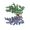
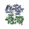
| Structure viewer | Molecule: MolmilJmol/JSmol |
|---|
- Downloads & links
-Download
| PDBx/mmCIF format | 2mad.cif.gz | 52.5 KB | Display | PDBx/mmCIF format |
|---|---|---|---|---|
| PDB format | pdb2mad.ent.gz | 34.8 KB | Display | PDB format |
| PDBx/mmJSON format | 2mad.json.gz | Tree view | PDBx/mmJSON format | |
| Others |  Other downloads Other downloads |
-Validation report
| Arichive directory | https://data.pdbj.org/pub/pdb/validation_reports/ma/2madftp://data.pdbj.org/pub/pdb/validation_reports/ma/2mad | HTTPS FTP |
|---|
-Related structure data


-Links
 PDBj
PDBj- Assembly
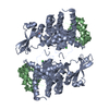
| Deposited unit | 
| ||||||||
|---|---|---|---|---|---|---|---|---|---|
| 1 | 
| ||||||||
| Unit cell |
| ||||||||
| Atom site foot note | 1: THE QUINONE CONTAINING TRYPTOPHAN (L 57).THE QUINONE OXYGEN IS GIVEN IN HETATM QTR. 2: LYS L 129 - ALA L 130 OMEGA ANGLE = 0.904 PEPTIDE BOND DEVIATES SIGNIFICANTLY FROM TRANS CONFORMATION | ||||||||
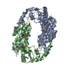
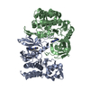
| Details | THE MOLECULE IS NORMALLY A TETRAMER. THE CRYSTALLOGRAPHIC ASYMMETRIC UNIT CONSISTS OF ONE-HALF OF THE TETRAMER, NAMELY ONE LIGHT CHAIN *L* AND ONE HEAVY CHAIN *H*. TO GENERATE THE FULL MOLECULE, THE FOLLOWING CRYSTALLOGRAPHIC TWO-FOLD OPERATION MUST BE APPLIED TO THE LIGHT AND HEAVY CHAINS PRESENTED IN THIS ENTRY -0.5 0.866025 0.0 0.0 0.866025 0.5 0.0 0.0 0.0 0.0 -1.0 208.368 |
-Components
| #1: Protein | Mass: 13668.154 Da / Num. of mol.: 1 Source method: isolated from a genetically manipulated source Source: (gene. exp.) Paracoccus versutus (bacteria) / References: UniProt: P22641, EC: 1.4.99.3 |
|---|---|
| #2: Protein | Mass: 38031.996 Da / Num. of mol.: 1 Source method: isolated from a genetically manipulated source Source: (gene. exp.) Paracoccus versutus (bacteria) / References: UniProt: P23006, EC: 1.4.99.3 |
| #3: Water | ChemComp-HOH /  Mass: 18.015 Da / Num. of mol.: 86 / Source method: isolated from a natural source / Formula: H2O Mass: 18.015 Da / Num. of mol.: 86 / Source method: isolated from a natural source / Formula: H2O |
| Compound details | THE L SUBUNIT CONTAINS THE SIDE CHAIN DERIVED COFACTOR TRYPTOPHYL TRYTOPHAN-QUINONE (MCINTYRE ET AL. ...THE L SUBUNIT CONTAINS THE SIDE CHAIN DERIVED COFACTOR TRYPTOPHYL |
| Sequence details | SEQUENCE ADVISORY NOTICE: DIFFERENCE BETWEEN SWISS-PROT AND PDB SEQUENCE. SWISS-PROT ENTRY NAME: ...SEQUENCE ADVISORY NOTICE: DIFFERENCE |
-Experimental details
-Experiment
| Experiment | Method: X-RAY DIFFRACTION |
|---|
- Sample preparation
| Crystal | Density Matthews: 4.9 Å3/Da / Density % sol: 74.92 % | |||||||||||||||||||||||||
|---|---|---|---|---|---|---|---|---|---|---|---|---|---|---|---|---|---|---|---|---|---|---|---|---|---|---|
| Crystal grow | *PLUS Temperature: 4 ℃ / pH: 5 / Method: vapor diffusion, hanging dropDetails: taken from Ubbink, M. et al (1991). Eur. J. Biochem., 202, 1003-1012. | |||||||||||||||||||||||||
| Components of the solutions | *PLUS
|
- Processing
| Software | Name: TNT / Classification: refinement | ||||||||||||||||||||||||||||||
|---|---|---|---|---|---|---|---|---|---|---|---|---|---|---|---|---|---|---|---|---|---|---|---|---|---|---|---|---|---|---|---|
| Refinement | Rfactor obs: 0.209 / Highest resolution: 2.25 Å | ||||||||||||||||||||||||||||||
| Refinement step | Cycle: LAST / Highest resolution: 2.25 Å
| ||||||||||||||||||||||||||||||
| Refine LS restraints |
| ||||||||||||||||||||||||||||||
| Software | *PLUS Name: TNT / Classification: refinement | ||||||||||||||||||||||||||||||
| Refinement | *PLUS Highest resolution: 2.25 Å / Rfactor obs: 0.209 | ||||||||||||||||||||||||||||||
| Solvent computation | *PLUS | ||||||||||||||||||||||||||||||
| Displacement parameters | *PLUS | ||||||||||||||||||||||||||||||
| Refine LS restraints | *PLUS
|
