 Movie
Movie Controller

[English] 日本語
 Yorodumi
- PDB-5ysb: Crystal structure of beta-1,2-glucooligosaccharide binding protei... -
+ Open data

- Basic information
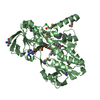
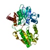
| Entry | Database: PDB / ID: 5ysb | ||||||
|---|---|---|---|---|---|---|---|
| Title | Crystal structure of beta-1,2-glucooligosaccharide binding protein in ligand-free form | ||||||
 Components Components | Lin1841 protein | ||||||
 Keywords Keywords | SUGAR BINDING PROTEIN / solute-binding protein / protein-carbohydrate complex / beta-1 / 2-glucooligosaccharide / sophorooligosaccharide / alpha/beta domain | ||||||
| Function / homology |  Function and homology information Function and homology informationmaltose binding / maltose transport / maltodextrin transmembrane transport / ATP-binding cassette (ABC) transporter complex, substrate-binding subunit-containing / metal ion binding Similarity search - Function | ||||||
| Biological species | Listeria innocua serovar 6a | ||||||
| Method |  X-RAY DIFFRACTION / SYNCHROTRON / MOLECULAR REPLACEMENT / Resolution: 2.2 Å X-RAY DIFFRACTION / SYNCHROTRON / MOLECULAR REPLACEMENT / Resolution: 2.2 Å | ||||||
 Authors Authors | Abe, K. / Nakajima, M. / Taguchi, H. / Arakawa, T. / Fushinobu, S. | ||||||
 Citation Citation | Journal: J. Biol. Chem. / Year: 2018 Title: Structural and thermodynamic insights into beta-1,2-glucooligosaccharide capture by a solute-binding protein inListeria innocua. Authors: Abe, K. / Sunagawa, N. / Terada, T. / Takahashi, Y. / Arakawa, T. / Igarashi, K. / Samejima, M. / Nakai, H. / Taguchi, H. / Nakajima, M. / Fushinobu, S. | ||||||
| History |
|
- Structure visualization
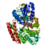
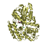
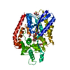
| Structure viewer | Molecule: MolmilJmol/JSmol |
|---|
- Downloads & links
-Download
| PDBx/mmCIF format | 5ysb.cif.gz | 315.2 KB | Display | PDBx/mmCIF format |
|---|---|---|---|---|
| PDB format | pdb5ysb.ent.gz | 254.3 KB | Display | PDB format |
| PDBx/mmJSON format | 5ysb.json.gz | Tree view | PDBx/mmJSON format | |
| Others |  Other downloads Other downloads |
-Validation report
| Arichive directory | https://data.pdbj.org/pub/pdb/validation_reports/ys/5ysbftp://data.pdbj.org/pub/pdb/validation_reports/ys/5ysb | HTTPS FTP |
|---|
-Related structure data
| Related structure data |  5ysdC  5yseC  5ysfC 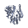 3uorS S: Starting model for refinement C: citing same article ( |
|---|---|
| Similar structure data |


-Links
 PDBj
PDBj


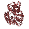
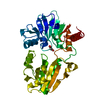
- Assembly
| Deposited unit | 
| ||||||||||||||||||
|---|---|---|---|---|---|---|---|---|---|---|---|---|---|---|---|---|---|---|---|
| 1 | 
| ||||||||||||||||||
| 2 | 
| ||||||||||||||||||
| Unit cell |
| ||||||||||||||||||
| Noncrystallographic symmetry (NCS) | NCS domain:
NCS domain segments: Component-ID: _ / Ens-ID: 1 / Beg auth comp-ID: VAL / Beg label comp-ID: VAL / End auth comp-ID: HIS / End label comp-ID: HIS / Refine code: _ / Auth seq-ID: 36 - 419 / Label seq-ID: 11 - 394
|
-Components
| #1: Protein | Mass: 44582.117 Da / Num. of mol.: 2 Source method: isolated from a genetically manipulated source Source: (gene. exp.)  Listeria innocua serovar 6a (strain ATCC BAA-680 / CLIP 11262) (bacteria) Listeria innocua serovar 6a (strain ATCC BAA-680 / CLIP 11262) (bacteria)Strain: ATCC BAA-680 / CLIP 11262 / Gene: lin1841 / Plasmid: pET30a / Production host: #2: Chemical | ChemComp-ZN /   Mass: 65.409 Da / Num. of mol.: 24 / Source method: obtained synthetically / Formula: Zn Mass: 65.409 Da / Num. of mol.: 24 / Source method: obtained synthetically / Formula: Zn#3: Chemical |   Mass: 106.120 Da / Num. of mol.: 2 / Source method: obtained synthetically / Formula: C4H10O3 Mass: 106.120 Da / Num. of mol.: 2 / Source method: obtained synthetically / Formula: C4H10O3#4: Water | ChemComp-HOH / |  Mass: 18.015 Da / Num. of mol.: 205 / Source method: isolated from a natural source / Formula: H2O Mass: 18.015 Da / Num. of mol.: 205 / Source method: isolated from a natural source / Formula: H2O |
|---|
-Experimental details
-Experiment
| Experiment | Method: X-RAY DIFFRACTION / Number of used crystals: 1 |
|---|
- Sample preparation
| Crystal | Density Matthews: 2.23 Å3/Da / Density % sol: 44.9 % |
|---|---|
| Crystal grow | Temperature: 298 K / Method: vapor diffusion, hanging drop / Details: 0.15M zinc acetate, 15% (w/v) PEG 3350 |
-Data collection
| Diffraction | Mean temperature: 100 K |
|---|---|
| Diffraction source | Source: SYNCHROTRON / Site: Photon Factory  / Beamline: AR-NW12A / Wavelength: 1 Å / Beamline: AR-NW12A / Wavelength: 1 Å |
| Detector | Type: ADSC QUANTUM 270 / Detector: CCD / Date: Jun 28, 2015 |
| Radiation | Monochromator: Numerical link type Si(111) double crystal / Protocol: SINGLE WAVELENGTH / Monochromatic (M) / Laue (L): M / Scattering type: x-ray |
| Radiation wavelength | Wavelength: 1 Å / Relative weight: 1 |
| Reflection | Resolution: 2.2→50 Å / Num. obs: 38141 / % possible obs: 98.8 % / Redundancy: 5.5 % / Rmerge(I) obs: 0.097 / Rpim(I) all: 0.047 / Net I/σ(I): 21 |
| Reflection shell | Resolution: 2.2→2.24 Å / Redundancy: 5.7 % / Rmerge(I) obs: 0.571 / Num. unique obs: 1912 / CC1/2: 0.906 / Rpim(I) all: 0.263 / % possible all: 100 |
- Processing
| Software |
| ||||||||||||||||||||||||||||||||||||||||||||||||||||||||||||||||||||||||||||||||||||||||||||||||||||||||||||||||||||||||||||||||||||||||||||||||||||||||||||||||||||||||||||||||||||||
|---|---|---|---|---|---|---|---|---|---|---|---|---|---|---|---|---|---|---|---|---|---|---|---|---|---|---|---|---|---|---|---|---|---|---|---|---|---|---|---|---|---|---|---|---|---|---|---|---|---|---|---|---|---|---|---|---|---|---|---|---|---|---|---|---|---|---|---|---|---|---|---|---|---|---|---|---|---|---|---|---|---|---|---|---|---|---|---|---|---|---|---|---|---|---|---|---|---|---|---|---|---|---|---|---|---|---|---|---|---|---|---|---|---|---|---|---|---|---|---|---|---|---|---|---|---|---|---|---|---|---|---|---|---|---|---|---|---|---|---|---|---|---|---|---|---|---|---|---|---|---|---|---|---|---|---|---|---|---|---|---|---|---|---|---|---|---|---|---|---|---|---|---|---|---|---|---|---|---|---|---|---|---|---|
| Refinement | Method to determine structure: MOLECULAR REPLACEMENT Starting model: 3UOR Resolution: 2.2→19.24 Å / Cor.coef. Fo:Fc: 0.952 / Cor.coef. Fo:Fc free: 0.922 / SU B: 10.858 / SU ML: 0.155 / Cross valid method: THROUGHOUT / ESU R: 0.32 / ESU R Free: 0.23 / Stereochemistry target values: MAXIMUM LIKELIHOOD / Details: HYDROGENS HAVE BEEN ADDED IN THE RIDING POSITIONS
| ||||||||||||||||||||||||||||||||||||||||||||||||||||||||||||||||||||||||||||||||||||||||||||||||||||||||||||||||||||||||||||||||||||||||||||||||||||||||||||||||||||||||||||||||||||||
| Solvent computation | Ion probe radii: 0.8 Å / Shrinkage radii: 0.8 Å / VDW probe radii: 1.2 Å / Solvent model: BABINET MODEL WITH MASK | ||||||||||||||||||||||||||||||||||||||||||||||||||||||||||||||||||||||||||||||||||||||||||||||||||||||||||||||||||||||||||||||||||||||||||||||||||||||||||||||||||||||||||||||||||||||
| Displacement parameters | Biso mean: 49.947 Å2
| ||||||||||||||||||||||||||||||||||||||||||||||||||||||||||||||||||||||||||||||||||||||||||||||||||||||||||||||||||||||||||||||||||||||||||||||||||||||||||||||||||||||||||||||||||||||
| Refinement step | Cycle: 1 / Resolution: 2.2→19.24 Å
| ||||||||||||||||||||||||||||||||||||||||||||||||||||||||||||||||||||||||||||||||||||||||||||||||||||||||||||||||||||||||||||||||||||||||||||||||||||||||||||||||||||||||||||||||||||||
| Refine LS restraints |
|