 Movie
Movie Controller

[English] 日本語
 Yorodumi
- PDB-1lwl: Crystal Structure of Cytochrome P450-cam with a Fluorescent Probe... -
+ Open data

- Basic information
| Entry | Database: PDB / ID: 1lwl | ||||||
|---|---|---|---|---|---|---|---|
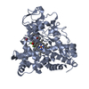
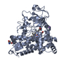
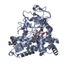
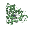
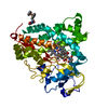
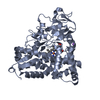
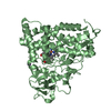
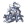
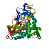
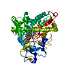
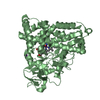
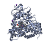
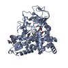
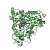
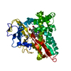
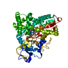
| Title | Crystal Structure of Cytochrome P450-cam with a Fluorescent Probe D-8-Ad (Adamantane-1-carboxylic acid-5-dimethylamino-naphthalene-1-sulfonylamino-octyl-amide) | ||||||
 Components Components | Cytochrome P450-cam | ||||||
 Keywords Keywords | OXIDOREDUCTASE / MONOOXYGENASE / ELECTRON TRANSFER / ENERGY TRANSFER / SUBSTRATE-BINDING / DANSYL / ADAMANTANE / Adamantane-1-carboxylic acid [4-(5-dimethylamino-naphthalene-1-sulfonylamino)-octyl]-amide / CHANNEL | ||||||
| Function / homology |  Function and homology information Function and homology informationcamphor 5-monooxygenase / camphor 5-monooxygenase activity / (+)-camphor catabolic process / iron ion binding / heme binding / cytoplasm Similarity search - Function | ||||||
| Biological species |  Pseudomonas putida (bacteria) Pseudomonas putida (bacteria) | ||||||
| Method |  X-RAY DIFFRACTION / MOLECULAR REPLACEMENT / Resolution: 2.2 Å X-RAY DIFFRACTION / MOLECULAR REPLACEMENT / Resolution: 2.2 Å | ||||||
 Authors Authors | Dunn, A.R. / Hays, A.M. / Goodin, D.B. / Stout, C.D. / Chiu, R. / Winkler, J.R. / Gray, H.B. | ||||||
 Citation Citation | Journal: J.AM.CHEM.SOC. / Year: 2002 Title: Fluorescent probes for cytochrome P450 structural characterization and inhibitor screening Authors: Dunn, A.R. / Hays, A.M. / Goodin, D.B. / Stout, C.D. / Chiu, R. / Winkler, J.R. / Gray, H.B. | ||||||
| History |
|
- Structure visualization
| Structure viewer | Molecule: MolmilJmol/JSmol |
|---|
- Downloads & links
-Download
| PDBx/mmCIF format | 1lwl.cif.gz | 101.2 KB | Display | PDBx/mmCIF format |
|---|---|---|---|---|
| PDB format | pdb1lwl.ent.gz | 76.2 KB | Display | PDB format |
| PDBx/mmJSON format | 1lwl.json.gz | Tree view | PDBx/mmJSON format | |
| Others |  Other downloads Other downloads |
-Validation report
| Arichive directory | https://data.pdbj.org/pub/pdb/validation_reports/lw/1lwlftp://data.pdbj.org/pub/pdb/validation_reports/lw/1lwl | HTTPS FTP |
|---|
-Related structure data
| Related structure data | 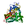 2cppS S: Starting model for refinement |
|---|---|
| Similar structure data |


-Links
 PDBj
PDBj
- Assembly
| Deposited unit | 
| ||||||||
|---|---|---|---|---|---|---|---|---|---|
| 1 |
| ||||||||
| Unit cell |
|
-Components
| #1: Protein | Mass: 46961.359 Da / Num. of mol.: 1 Source method: isolated from a genetically manipulated source Source: (gene. exp.) Pseudomonas putida (bacteria) / Plasmid: PUS200 / Production host: |
|---|---|
| #2: Chemical | ChemComp-HEM /   Mass: 616.487 Da / Num. of mol.: 1 / Source method: obtained synthetically / Formula: C34H32FeN4O4 Mass: 616.487 Da / Num. of mol.: 1 / Source method: obtained synthetically / Formula: C34H32FeN4O4 |
| #3: Chemical | ChemComp-DSO /   Mass: 539.772 Da / Num. of mol.: 1 / Source method: obtained synthetically / Formula: C31H45N3O3S Mass: 539.772 Da / Num. of mol.: 1 / Source method: obtained synthetically / Formula: C31H45N3O3S |
| #4: Water | ChemComp-HOH /  Mass: 18.015 Da / Num. of mol.: 302 / Source method: isolated from a natural source / Formula: H2O Mass: 18.015 Da / Num. of mol.: 302 / Source method: isolated from a natural source / Formula: H2O |
-Experimental details
-Experiment
| Experiment | Method: X-RAY DIFFRACTION / Number of used crystals: 1 |
|---|
- Sample preparation
| Crystal | Density Matthews: 2.42 Å3/Da / Density % sol: 49.28 % | ||||||||||||||||||||||||||||
|---|---|---|---|---|---|---|---|---|---|---|---|---|---|---|---|---|---|---|---|---|---|---|---|---|---|---|---|---|---|
| Crystal grow | Temperature: 277 K / Method: vapor diffusion, hanging drop / pH: 5.5 Details: Citrate, PEG, KCl, DTT, pH 5.5, VAPOR DIFFUSION, HANGING DROP, temperature 277K | ||||||||||||||||||||||||||||
| Crystal grow | *PLUS Temperature: 4 ℃ | ||||||||||||||||||||||||||||
| Components of the solutions | *PLUS
|
-Data collection
| Diffraction | Mean temperature: 105 K |
|---|---|
| Diffraction source | Source: ROTATING ANODE / Type: RIGAKU RU200 / Wavelength: 1.5418 Å |
| Detector | Type: RIGAKU RAXIS IV / Detector: IMAGE PLATE / Date: Jan 16, 2002 / Details: osmic confocal mirrors |
| Radiation | Monochromator: graphite / Protocol: SINGLE WAVELENGTH / Monochromatic (M) / Laue (L): M / Scattering type: x-ray |
| Radiation wavelength | Wavelength: 1.5418 Å / Relative weight: 1 |
| Reflection | Resolution: 2.2→20 Å / Num. all: 21045 / Num. obs: 21045 / % possible obs: 93.3 % / Observed criterion σ(F): 1 / Observed criterion σ(I): -3 / Redundancy: 5.2 % / Biso Wilson estimate: 26.2 Å2 / Rsym value: 0.102 / Net I/σ(I): 13.9 |
| Reflection shell | Resolution: 2.2→2.3 Å / Mean I/σ(I) obs: 2.5 / Rsym value: 0.266 / % possible all: 63.8 |
| Reflection | *PLUS Lowest resolution: 20 Å / Num. measured all: 115720 / Rmerge(I) obs: 0.102 |
| Reflection shell | *PLUS % possible obs: 63.8 % / Rmerge(I) obs: 0.266 |
- Processing
| Software |
| |||||||||||||||||||||||||
|---|---|---|---|---|---|---|---|---|---|---|---|---|---|---|---|---|---|---|---|---|---|---|---|---|---|---|
| Refinement | Method to determine structure: MOLECULAR REPLACEMENT Starting model: PDB ENTRY 2CPP Resolution: 2.2→20 Å / σ(F): 0 / Stereochemistry target values: Engh & Huber
| |||||||||||||||||||||||||
| Refinement step | Cycle: LAST / Resolution: 2.2→20 Å
| |||||||||||||||||||||||||
| LS refinement shell | Resolution: 2.2→2.3 Å
| |||||||||||||||||||||||||
| Refinement | *PLUS Lowest resolution: 20 Å / % reflection Rfree: 4.8 % | |||||||||||||||||||||||||
| Solvent computation | *PLUS | |||||||||||||||||||||||||
| Displacement parameters | *PLUS | |||||||||||||||||||||||||
| Refine LS restraints | *PLUS
| |||||||||||||||||||||||||
| LS refinement shell | *PLUS Highest resolution: 2.2 Å / Lowest resolution: 2.3 Å / Rfactor Rfree: 0.33 / Rfactor Rwork: 0.285 |
