| 登録情報 | データベース: PDB / ID: 1iep
|
|---|
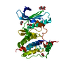
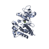
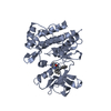
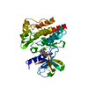
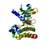
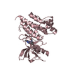
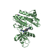
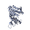
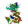
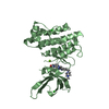
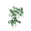
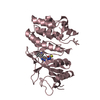
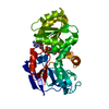
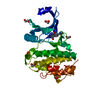
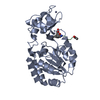
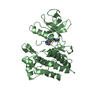
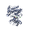
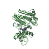
| タイトル | CRYSTAL STRUCTURE OF THE C-ABL KINASE DOMAIN IN COMPLEX WITH STI-571. |
|---|
 要素 要素 | PROTO-ONCOGENE TYROSINE-PROTEIN KINASE ABL |
|---|
 キーワード キーワード | TRANSFERASE / KINASE / KINASE INHIBITOR / STI-571 / ACTIVATION LOOP |
|---|
| 機能・相同性 |  機能・相同性情報 機能・相同性情報
Role of ABL in ROBO-SLIT signaling / HDR through Single Strand Annealing (SSA) / RHO GTPases Activate WASPs and WAVEs / Cyclin D associated events in G1 / MLL4 and MLL3 complexes regulate expression of PPARG target genes in adipogenesis and hepatic steatosis / Recruitment and ATM-mediated phosphorylation of repair and signaling proteins at DNA double strand breaks / Turbulent (oscillatory, disturbed) flow shear stress activates signaling by PIEZO1 and integrins in endothelial cells / protein localization to cytoplasmic microtubule plus-end / DNA conformation change / DN4 thymocyte differentiation ...Role of ABL in ROBO-SLIT signaling / HDR through Single Strand Annealing (SSA) / RHO GTPases Activate WASPs and WAVEs / Cyclin D associated events in G1 / MLL4 and MLL3 complexes regulate expression of PPARG target genes in adipogenesis and hepatic steatosis / Recruitment and ATM-mediated phosphorylation of repair and signaling proteins at DNA double strand breaks / Turbulent (oscillatory, disturbed) flow shear stress activates signaling by PIEZO1 and integrins in endothelial cells / protein localization to cytoplasmic microtubule plus-end / DNA conformation change / DN4 thymocyte differentiation / response to epinephrine / phospholipase C-inhibiting G protein-coupled receptor signaling pathway / podocyte apoptotic process / RUNX1 regulates transcription of genes involved in differentiation of HSCs / delta-catenin binding / transitional one stage B cell differentiation / regulation of postsynaptic specialization assembly / regulation of modification of synaptic structure / cerebellum morphogenesis / regulation of cellular senescence / neuroepithelial cell differentiation / B cell proliferation involved in immune response / Regulation of actin dynamics for phagocytic cup formation / positive regulation of extracellular matrix organization / positive regulation of Wnt signaling pathway, planar cell polarity pathway / microspike assembly / B-1 B cell homeostasis / neuropilin signaling pathway / neuropilin binding / regulation of extracellular matrix organization / Myogenesis / bubble DNA binding / positive regulation of establishment of T cell polarity / activated T cell proliferation / positive regulation of blood vessel branching / proline-rich region binding / negative regulation of mitotic cell cycle / regulation of Cdc42 protein signal transduction / circulatory system development / mitogen-activated protein kinase binding / positive regulation of dendrite development / syntaxin binding / positive regulation of cell migration involved in sprouting angiogenesis / alpha-beta T cell differentiation / positive regulation of peptidyl-tyrosine phosphorylation / regulation of axon extension / regulation of T cell differentiation / negative regulation of cell-cell adhesion / neuromuscular process controlling balance / positive regulation of osteoblast proliferation / platelet-derived growth factor receptor-beta signaling pathway / platelet-derived growth factor receptor signaling pathway / positive regulation of vasoconstriction / cell leading edge / Bergmann glial cell differentiation / B cell proliferation / regulation of microtubule polymerization / myoblast proliferation / negative regulation of long-term synaptic potentiation / negative regulation of cellular senescence / associative learning / signal transduction in response to DNA damage / positive regulation of focal adhesion assembly / negative regulation of BMP signaling pathway / canonical NF-kappaB signal transduction / cardiac muscle cell proliferation / ephrin receptor signaling pathway / phagocytosis / BMP signaling pathway / cellular response to transforming growth factor beta stimulus / negative regulation of endothelial cell apoptotic process / positive regulation of T cell migration / endothelial cell migration / negative regulation of double-strand break repair via homologous recombination / spleen development / ephrin receptor binding / four-way junction DNA binding / positive regulation of stress fiber assembly / ruffle / ERK1 and ERK2 cascade / phosphotyrosine residue binding / actin filament polymerization / positive regulation of substrate adhesion-dependent cell spreading / substrate adhesion-dependent cell spreading / positive regulation of mitotic cell cycle / positive regulation of interleukin-2 production / SH2 domain binding / peptidyl-tyrosine phosphorylation / thymus development / protein kinase C binding / response to endoplasmic reticulum stress / positive regulation of release of sequestered calcium ion into cytosol / integrin-mediated signaling pathway / post-embryonic development / B cell receptor signaling pathway / regulation of actin cytoskeleton organization / non-membrane spanning protein tyrosine kinase activity / non-specific protein-tyrosine kinase / neural tube closure / establishment of localization in cell類似検索 - 分子機能 F-actin binding / F-actin binding / F-actin binding domain (FABD) / Tyrosine-protein kinase ABL, SH2 domain / : / SH3 domain / SH2 domain / Src homology 2 (SH2) domain profile. / Src homology 2 domains / SH2 domain ...F-actin binding / F-actin binding / F-actin binding domain (FABD) / Tyrosine-protein kinase ABL, SH2 domain / : / SH3 domain / SH2 domain / Src homology 2 (SH2) domain profile. / Src homology 2 domains / SH2 domain / Src homology 3 domains / SH2 domain superfamily / SH3-like domain superfamily / Src homology 3 (SH3) domain profile. / SH3 domain / Tyrosine-protein kinase, catalytic domain / Tyrosine kinase, catalytic domain / Tyrosine protein kinases specific active-site signature. / Tyrosine-protein kinase, active site / Serine-threonine/tyrosine-protein kinase, catalytic domain / Protein tyrosine and serine/threonine kinase / Phosphorylase Kinase; domain 1 / Phosphorylase Kinase; domain 1 / Transferase(Phosphotransferase) domain 1 / Transferase(Phosphotransferase); domain 1 / Protein kinase, ATP binding site / Protein kinases ATP-binding region signature. / Protein kinase domain profile. / Protein kinase domain / Protein kinase-like domain superfamily / 2-Layer Sandwich / Orthogonal Bundle / Mainly Alpha / Alpha Beta類似検索 - ドメイン・相同性 |
|---|
| 生物種 |   Mus musculus (ハツカネズミ) Mus musculus (ハツカネズミ) |
|---|
| 手法 |  X線回折 / シンクロトロン / フーリエ合成 / 解像度: 2.1 Å X線回折 / シンクロトロン / フーリエ合成 / 解像度: 2.1 Å |
|---|
 データ登録者 データ登録者 | Nagar, B. / Bornmann, W. / Schindler, T. / Clarkson, B. / Kuriyan, J. |
|---|
 引用 引用 | ジャーナル: Cancer Res. / 年: 2002
タイトル: Crystal structures of the kinase domain of c-Abl in complex with the small molecule inhibitors PD173955 and imatinib (STI-571)
著者: Nagar, B. / Bornmann, W. / Pellicena, P. / Schindler, T. / Veach, D.R. / Miller, W.T. / Clarkson, B. / Kuriyan, J. |
|---|
| 履歴 | | 登録 | 2001年4月10日 | 登録サイト: RCSB / 処理サイト: RCSB |
|---|
| 改定 1.0 | 2001年4月18日 | Provider: repository / タイプ: Initial release |
|---|
| 改定 1.1 | 2008年4月27日 | Group: Version format compliance |
|---|
| 改定 1.2 | 2011年7月13日 | Group: Version format compliance |
|---|
| 改定 1.3 | 2023年8月9日 | Group: Data collection / Database references ...Data collection / Database references / Derived calculations / Refinement description / Structure summary
カテゴリ: chem_comp / chem_comp_atom ...chem_comp / chem_comp_atom / chem_comp_bond / database_2 / pdbx_initial_refinement_model / struct_ref_seq_dif / struct_site
Item: _chem_comp.pdbx_synonyms / _database_2.pdbx_DOI ..._chem_comp.pdbx_synonyms / _database_2.pdbx_DOI / _database_2.pdbx_database_accession / _struct_ref_seq_dif.details / _struct_site.pdbx_auth_asym_id / _struct_site.pdbx_auth_comp_id / _struct_site.pdbx_auth_seq_id |
|---|
|
|---|
 ムービー
ムービー コントローラー


 データを開く
 基本情報
基本情報 構造の表示
構造の表示 ダウンロードとリンク
 その他のダウンロード


 PDBj
PDBj


 集合体
集合体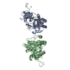

 Spodoptera frugiperda (ツマジロクサヨトウ)
 分子量: 35.453 Da / 分子数: 6 / 由来タイプ: 合成 / 式: Cl
 分子量: 493.603 Da / 分子数: 2 / 由来タイプ: 合成 / 式: C29H31N7O / コメント: 薬剤, 阻害剤*YM
 分子量: 18.015 Da / 分子数: 172 / 由来タイプ: 天然 / 式: H2O
 試料調製
試料調製 / ビームライン: F1 / 波長: 0.949 Å
 解析
解析