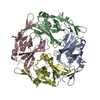
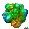
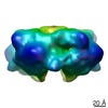
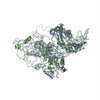
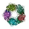
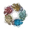
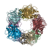
ジャーナル: Biochim Biophys Acta / 年: 2011 タイトル: Self-directed assembly and clustering of the cytoplasmic domains of inwardly rectifying Kir2.1 potassium channels on association with PSD-95. 著者: Svetlana Fomina / Tina D Howard / Olivia K Sleator / Marina Golovanova / Liam O'Ryan / Mark L Leyland / J Günter Grossmann / Richard F Collins / Stephen M Prince / 要旨: The interaction of the extra-membranous domain of tetrameric inwardly rectifying Kir2.1 ion channels (Kir2.1NC(4)) with the membrane associated guanylate kinase protein PSD-95 has been studied using ...The interaction of the extra-membranous domain of tetrameric inwardly rectifying Kir2.1 ion channels (Kir2.1NC(4)) with the membrane associated guanylate kinase protein PSD-95 has been studied using Transmission Electron Microscopy in negative stain. Three types of complexes were observed in electron micrographs corresponding to a 1:1 complex, a large self-enclosed tetrad complex and extended chains of linked channel domains. Using models derived from small angle X-ray scattering experiments in which high resolution structures from X-ray crystallographic and Nuclear Magnetic Resonance studies are positioned, the envelopes from single particle analysis can be resolved as a Kir2.1NC(4):PSD-95 complex and a tetrad of this unit (Kir2.1NC(4):PSD-95)(4). The tetrad complex shows the close association of the Kir2.1 cytoplasmic domains and the influence of PSD-95 mediated self-assembly on the clustering of these channels.
タイプ: NEGATIVE 詳細: Samples were adsorbed onto freshly glow discharged carbon film grids. Sample solution was pipetted into the grid followed by blotting, de-ionized water was then applied for 10s followed by ...詳細: Samples were adsorbed onto freshly glow discharged carbon film grids. Sample solution was pipetted into the grid followed by blotting, de-ionized water was then applied for 10s followed by blotting, 2% w/v Uranyl Acetate solution was applied followed by a final blotting step.
グリッド
詳細: 400 mesh Copper
凍結
凍結剤: NONE / 装置: OTHER
-
電子顕微鏡法
顕微鏡
FEI TECNAI 10
詳細
Low dose
撮影
カテゴリ: FILM フィルム・検出器のモデル: GATAN ORIUS SC200 (2k x 2k) デジタル化 - スキャナー: OTHER / デジタル化 - サンプリング間隔: 10 µm / 実像数: 22 / ビット/ピクセル: 8
Particles initially selected using automated particle picking based on a subset of representative particles on a single micrograph, followed by model-based picking.
CTF補正
詳細: Parameters determined using Scattering curve
最終 再構成
想定した対称性 - 点群: C4 (4回回転対称) / 解像度のタイプ: BY AUTHOR / 解像度: 17.2 Å / 解像度の算出法: FSC 0.5 CUT-OFF / ソフトウェア - 名称: EMAN / 使用した粒子像数: 49012
 ムービー
ムービー コントローラー


 データを開く
 基本情報
基本情報 マップデータ
 試料
試料 キーワード
キーワード 機能・相同性情報
 データ登録者
 引用
引用
 構造の表示
構造の表示 UCSF Chimera
 ダウンロードとリンク
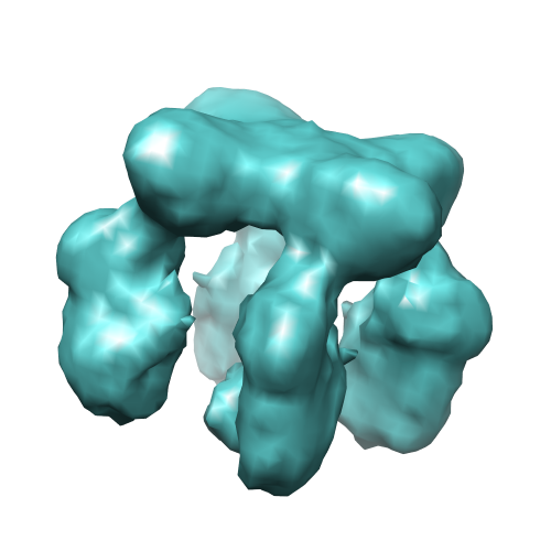 image1764.png
 http://ftp.pdbj.org/pub/emdb/structures/EMD-1764
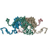


 試料の構成要素
 解析
解析 電子顕微鏡法