 Movie
Movie Controller


+ Open data

- Basic information
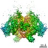
| Entry | Database: EMDB / ID: EMD-13591 | |||||||||
|---|---|---|---|---|---|---|---|---|---|---|
| Title | Structure of SidJ/CaM bound to SdeA in post-catalysis state | |||||||||
 Map data Map data | ||||||||||
 Sample Sample |
| |||||||||
| Function / homology |  Function and homology information Function and homology informationLigases / NAD+-protein-arginine ADP-ribosyltransferase / negative regulation of calcium ion transmembrane transporter activity / deNEDDylase activity / NAD+-protein-arginine ADP-ribosyltransferase activity / protein deneddylation / Transferases; Acyltransferases; Aminoacyltransferases / K63-linked deubiquitinase activity / positive regulation of cyclic-nucleotide phosphodiesterase activity / negative regulation of calcium ion export across plasma membrane ...Ligases / NAD+-protein-arginine ADP-ribosyltransferase / negative regulation of calcium ion transmembrane transporter activity / deNEDDylase activity / NAD+-protein-arginine ADP-ribosyltransferase activity / protein deneddylation / Transferases; Acyltransferases; Aminoacyltransferases / K63-linked deubiquitinase activity / positive regulation of cyclic-nucleotide phosphodiesterase activity / negative regulation of calcium ion export across plasma membrane / positive regulation of ryanodine-sensitive calcium-release channel activity / regulation of cell communication by electrical coupling involved in cardiac conduction / protein deubiquitination / negative regulation of peptidyl-threonine phosphorylation / protein phosphatase activator activity / ligase activity / positive regulation of phosphoprotein phosphatase activity / adenylate cyclase binding / catalytic complex / detection of calcium ion / regulation of cardiac muscle contraction / negative regulation of ryanodine-sensitive calcium-release channel activity / calcium channel inhibitor activity / regulation of cardiac muscle contraction by regulation of the release of sequestered calcium ion / regulation of release of sequestered calcium ion into cytosol by sarcoplasmic reticulum / cysteine-type peptidase activity / positive regulation of protein dephosphorylation / regulation of calcium-mediated signaling / titin binding / positive regulation of protein autophosphorylation / voltage-gated potassium channel complex / sperm midpiece / calcium channel complex / substantia nigra development / adenylate cyclase activator activity / regulation of heart rate / nucleotidyltransferase activity / sarcomere / protein serine/threonine kinase activator activity / positive regulation of peptidyl-threonine phosphorylation / regulation of cytokinesis / positive regulation of protein serine/threonine kinase activity / spindle microtubule / spindle pole / response to calcium ion / G2/M transition of mitotic cell cycle / calcium-dependent protein binding / host cell / myelin sheath / transferase activity / vesicle / transmembrane transporter binding / Hydrolases; Acting on peptide bonds (peptidases); Cysteine endopeptidases / protein ubiquitination / G protein-coupled receptor signaling pathway / nucleotide binding / centrosome / calcium ion binding / protein kinase binding / protein-containing complex / proteolysis / extracellular region / membrane / nucleus / metal ion binding / plasma membrane / cytoplasm Similarity search - Function | |||||||||
| Biological species |   Legionella pneumophila (bacteria) / Legionella pneumophila (bacteria) /  Homo sapiens (human) Homo sapiens (human) | |||||||||
| Method | single particle reconstruction / cryo EM / Resolution: 3.7 Å | |||||||||
 Authors Authors | Adams M / Bhogaraju S | |||||||||
| Funding support | European Union, 1 items
| |||||||||
 Citation Citation | Journal: Nat Commun / Year: 2021 Title: Structural basis for protein glutamylation by the Legionella pseudokinase SidJ. Authors: Michael Adams / Rahul Sharma / Thomas Colby / Felix Weis / Ivan Matic / Sagar Bhogaraju /   Abstract: Legionella pneumophila (LP) avoids phagocytosis by secreting nearly 300 effector proteins into the host cytosol. SidE family of effectors (SdeA, SdeB, SdeC and SidE) employ phosphoribosyl ...Legionella pneumophila (LP) avoids phagocytosis by secreting nearly 300 effector proteins into the host cytosol. SidE family of effectors (SdeA, SdeB, SdeC and SidE) employ phosphoribosyl ubiquitination to target multiple host Rab GTPases and innate immune factors. To suppress the deleterious toxicity of SidE enzymes in a timely manner, LP employs a metaeffector named SidJ. Upon activation by host Calmodulin (CaM), SidJ executes an ATP-dependent glutamylation to modify the catalytic residue Glu860 in the mono-ADP-ribosyl transferase (mART) domain of SdeA. SidJ is a unique glutamylase that adopts a kinase-like fold but contains two nucleotide-binding pockets. There is a lack of consensus about the substrate recognition and catalytic mechanism of SidJ. Here, we determined the cryo-EM structure of SidJ in complex with its substrate SdeA in two different states of catalysis. Our structures reveal that both phosphodiesterase (PDE) and mART domains of SdeA make extensive contacts with SidJ. In the pre-glutamylation state structure of the SidJ-SdeA complex, adenylylated E860 of SdeA is inserted into the non-canonical (migrated) nucleotide-binding pocket of SidJ. Structure-based mutational analysis indicates that SidJ employs its migrated pocket for the glutamylation of SdeA. Finally, using mass spectrometry, we identified several transient autoAMPylation sites close to both the catalytic pockets of SidJ. Our data provide unique insights into the substrate recognition and the mechanism of protein glutamylation by the pseudokinase SidJ. #1: Journal: Acta Crystallogr., Sect. D: Biol. Crystallogr. / Year: 2010Title: PHENIX: a comprehensive Python-based system for macromolecular structure solution Authors: Adams PD / Afonine PV / Bunkoczi G / Chen VB / Davis IW / Echols N / Headd JJ / Hung L / Kapral GJ / Grosse RW / McCoy AJ / Moriarty NW / Oeffner R / Read RJ / Richardson DC / Richardson JS ...Authors: Adams PD / Afonine PV / Bunkoczi G / Chen VB / Davis IW / Echols N / Headd JJ / Hung L / Kapral GJ / Grosse RW / McCoy AJ / Moriarty NW / Oeffner R / Read RJ / Richardson DC / Richardson JS / Terwilliger TC / Zwart PH | |||||||||
| History |
|
- Structure visualization
| Movie |
Movie viewer |
|---|---|
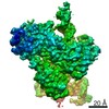
| Structure viewer | EM map: SurfViewMolmilJmol/JSmol |
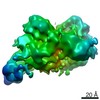
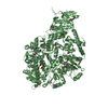
| Supplemental images |

- Downloads & links
-EMDB archive
| Map data | emd_13591.map.gz | 75.8 MB | EMDB map data format | |
|---|---|---|---|---|
| Header (meta data) | emd-13591-v30.xmlemd-13591.xml | 16.9 KB 16.9 KB | Display Display | EMDB header |
| FSC (resolution estimation) | emd_13591_fsc.xml | 11.4 KB | Display | FSC data file |
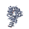
| Images | 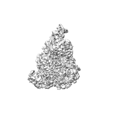 emd_13591.png emd_13591.png | 48.3 KB | ||
| Masks | emd_13591_msk_1.map | 125 MB | Mask map | |
| Archive directory |  http://ftp.pdbj.org/pub/emdb/structures/EMD-13591ftp://ftp.pdbj.org/pub/emdb/structures/EMD-13591 http://ftp.pdbj.org/pub/emdb/structures/EMD-13591ftp://ftp.pdbj.org/pub/emdb/structures/EMD-13591 | HTTPS FTP |
-Validation report
| Summary document | emd_13591_validation.pdf.gz | 349.4 KB | Display | EMDB validaton report |
|---|---|---|---|---|
| Full document | emd_13591_full_validation.pdf.gz | 348.9 KB | Display | |
| Data in XML | emd_13591_validation.xml.gz | 12 KB | Display | |
| Data in CIF | emd_13591_validation.cif.gz | 15.8 KB | Display | |
| Arichive directory | https://ftp.pdbj.org/pub/emdb/validation_reports/EMD-13591ftp://ftp.pdbj.org/pub/emdb/validation_reports/EMD-13591 | HTTPS FTP |
-Related structure data
| Related structure data | 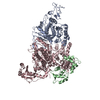 7pqeMC  7ppoC C: citing same article ( M: atomic model generated by this map |
|---|---|
| Similar structure data |


-Links
| EMDB pages | EMDB (EBI/PDBe) / EMDataResource |
|---|---|
| Related items in Molecule of the Month |

-Map
| File | Download / File: emd_13591.map.gz / Format: CCP4 / Size: 125 MB / Type: IMAGE STORED AS FLOATING POINT NUMBER (4 BYTES) | ||||||||||||||||||||||||||||||||||||||||||||||||||||||||||||||||||||
|---|---|---|---|---|---|---|---|---|---|---|---|---|---|---|---|---|---|---|---|---|---|---|---|---|---|---|---|---|---|---|---|---|---|---|---|---|---|---|---|---|---|---|---|---|---|---|---|---|---|---|---|---|---|---|---|---|---|---|---|---|---|---|---|---|---|---|---|---|---|
| Voxel size | X=Y=Z: 0.941 Å | ||||||||||||||||||||||||||||||||||||||||||||||||||||||||||||||||||||
| Density |
| ||||||||||||||||||||||||||||||||||||||||||||||||||||||||||||||||||||
| Symmetry | Space group: 1 | ||||||||||||||||||||||||||||||||||||||||||||||||||||||||||||||||||||
| Details | EMDB XML:
CCP4 map header:
| ||||||||||||||||||||||||||||||||||||||||||||||||||||||||||||||||||||
-Supplemental data
-Mask #1
| File | emd_13591_msk_1.map | ||||||||||||
|---|---|---|---|---|---|---|---|---|---|---|---|---|---|
| Projections & Slices |
| ||||||||||||
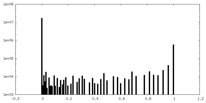
| Density Histograms |
 Z
Z Y
Y X
X


- Sample components
-Entire : SidJ/CaM-SdeA
| Entire | Name: SidJ/CaM-SdeA |
|---|---|
| Components |
|
-Supramolecule #1: SidJ/CaM-SdeA
| Supramolecule | Name: SidJ/CaM-SdeA / type: complex / ID: 1 / Parent: 0 / Macromolecule list: #1-#3 |
|---|
-Supramolecule #2: Septation initiation protein and SidJ
| Supramolecule | Name: Septation initiation protein and SidJ / type: complex / ID: 2 / Parent: 1 / Macromolecule list: #1-#2 |
|---|---|
| Source (natural) | Organism: Legionella pneumophila (bacteria) |
| Recombinant expression | Organism: |
-Supramolecule #3: Calmodulin
| Supramolecule | Name: Calmodulin / type: complex / ID: 3 / Parent: 1 / Macromolecule list: #3 |
|---|---|
| Source (natural) | Organism: Homo sapiens (human) |
| Recombinant expression | Organism: |
-Macromolecule #1: Ubiquitinating/deubiquitinating enzyme SdeA
| Macromolecule | Name: Ubiquitinating/deubiquitinating enzyme SdeA / type: protein_or_peptide / ID: 1 / Number of copies: 1 / Enantiomer: LEVO EC number: Hydrolases; Acting on peptide bonds (peptidases); Cysteine endopeptidases |
|---|---|
| Source (natural) | Organism: Legionella pneumophila (bacteria) |
| Molecular weight | Theoretical: 110.875203 KDa |
| Recombinant expression | Organism: |
| Sequence | String: HHHHHHSAGL EVLFQGPMVG FSLYTDDTVK AAAQYAYDNY LGKPYTGSVE SAPANFGGRM VYRQHHGLSH TLRTMAYAEL IVEEARKAK LRGETLGKFK DGRTIADVTP QELKKIMIAQ AFFVAGRDDE ASDAKNYQKY HEQSRDAFLK YVKDNESTLI P DVFKDQED ...String: HHHHHHSAGL EVLFQGPMVG FSLYTDDTVK AAAQYAYDNY LGKPYTGSVE SAPANFGGRM VYRQHHGLSH TLRTMAYAEL IVEEARKAK LRGETLGKFK DGRTIADVTP QELKKIMIAQ AFFVAGRDDE ASDAKNYQKY HEQSRDAFLK YVKDNESTLI P DVFKDQED VNFYARVIED KSHDWESTPA HVLINQGHMV DLVRVKQPPE SFLQRYFSSM QRWIGSQATE AVFGIQRQFF HA TYEVVAG FDSDNKEPHL VVSGLGRYVI GEDGQPIREA PKKGQKEGDL KVFPQTYKLK ENERLMRVDE FLKLPEIQNT FPG SGKHLQ GGMPGMNEMD YWNRLNSLNR ARCENDVDFC LKQLQTAHDK AKIEPIKQAF QSSKGKERRQ PNVDEIAAAR IIQQ ILANP DCIHDDHVLI NGQKLEQQFF RDLLAKCEMA VVGSLLNDTD IGNIDTLMRH EKDTEFHSTN PEAVPVKIGE YWIND QRIN NSSGNITQKK HDLIFLMQND AWYFSRVNAI AQNRDKGSTF KEVLITTLMT PLTSKALVDT SQAKPPTRLF RGLNLS EEF TKGLIDQANA MIANTTERLF TDHSPEAFKQ IKLNDLSKMS GRTNASTTTE IKLVKETWDS NVIFEMLDPD GLLHSKQ VG RHGEGTESEF SVYLPEDVAL VPVKVTLDGK TQKGENRYVF TFVAVKSPDF TPRHESGYAV EPFLRMQAAK LAEVKSSI E KAQRAPDLET IFNLQNEVEA VQYSHLSTGY KNFLKNTVGP VLENSLSGLM ESDTDTLSKA LAAFPSDTQW SAFNFEEAR QAKRQMDAIK QMVGNKVVLD ALTQCQDALE KQNIAGALDA LKKIPSEKEM GTIRRELREQ IQSARQELES LQRAVVTPVV TDEKKVRER YDALIENTSK KITELETGKL PNLDAVKKGI SNLSNLKQEV TVLRNEKIRM HVGTDKVDFS DVEKLEQQIQ V IDTKLADA YLLEVTKQIS A |
-Macromolecule #2: Calmodulin-dependent glutamylase SidJ
| Macromolecule | Name: Calmodulin-dependent glutamylase SidJ / type: protein_or_peptide / ID: 2 / Number of copies: 1 / Enantiomer: LEVO / EC number: Ligases |
|---|---|
| Source (natural) | Organism: Legionella pneumophila (bacteria) |
| Molecular weight | Theoretical: 91.826992 KDa |
| Recombinant expression | Organism: |
| Sequence | String: HHHHHHSAGL EVLFQGPMVK QYYFARRGET STHDTSLPPP VKVLSGRSIP LKEIPFETTR NELVQIYLTS VDQLIKSNKL NSIPSQQIA SHYLFLRSLA NSETDGIKKN QILSLAKPLG IYLASKEPHV WKTINELIEK SEYPIIHYLK NNRAHSNFML A LIHEYHKE ...String: HHHHHHSAGL EVLFQGPMVK QYYFARRGET STHDTSLPPP VKVLSGRSIP LKEIPFETTR NELVQIYLTS VDQLIKSNKL NSIPSQQIA SHYLFLRSLA NSETDGIKKN QILSLAKPLG IYLASKEPHV WKTINELIEK SEYPIIHYLK NNRAHSNFML A LIHEYHKE PLTKNQSAFV QKFRDSSVFL FPNPIYTAWL AHSYDEDSSF NPMFRERLST NFYHSTLTDN LLLRTEPKEV TL SSEHHYK KEKGPIDSSF RYQMSSDRLL RIQGRTLLFS TPQNDVVAVK VQKRGEPKST LEEEFQMADY LLKHQSRLDV YSK LPQPLG QYSVKKSEIL EISRGSLDFE RFKTLIGDSK DLEVYVYKAP LTYFTYLHDK NQDLEDLTAS VKTNVHDLFV LLRE GIMFP QLADIFHTHF GEDEREDKGR YQALVQLLNV LQFQLGRIDK WQKAVEYVNL RSSGLADLGD SLPITSLFTS SDFTK HYFS ALLTGGYHPT FFDKSSGTAN SLFTGKRRLF GNYLYLNTIA EYLLVIQLTL GSYGDKVTRD MMDKPKKEAV WRELAN VMF TSCAEAIHIM TGIPQSRALT LLKQRANIEK HFRQTQFWMT PDYSKLDEDT LQMEQYSIYS GEPEYEFTDK LVSGVGL SV DGTHQDLGGY NRESPLRELE KLLYATVTLI EGTMQLDKEF FKQLQQVEKI LSGEIKTDAN SCFEAVAQLL DLARPRCH F QKRLVLSYYE EAKLKYPSAP TDAYDSRFQV VAKTNAAITI QRFWRETRKN LSENSDIESE KPESERTTDK RLK |
-Macromolecule #3: Calmodulin
| Macromolecule | Name: Calmodulin / type: protein_or_peptide / ID: 3 / Number of copies: 1 / Enantiomer: LEVO |
|---|---|
| Source (natural) | Organism: Homo sapiens (human) |
| Molecular weight | Theoretical: 19.066006 KDa |
| Recombinant expression | Organism: |
| Sequence | String: HHHHHHSSGL EVLFQGPHMM ADQLTEEQIA EFKEAFSLFD KDGDGTITTK ELGTVMRSLG QNPTEAELQD MINEVDADGN GTIDFPEFL TMMARKMKDT DSEEEIREAF RVFDKDGNGY ISAAELRHVM TNLGEKLTDE EVDEMIREAD IDGDGQVNYE E FVQMMTAK |
-Macromolecule #4: MAGNESIUM ION
| Macromolecule | Name: MAGNESIUM ION / type: ligand / ID: 4 / Number of copies: 1 / Formula: MG |
|---|---|
| Molecular weight | Theoretical: 24.305 Da |
-Macromolecule #5: CALCIUM ION
| Macromolecule | Name: CALCIUM ION / type: ligand / ID: 5 / Number of copies: 1 / Formula: CA |
|---|---|
| Molecular weight | Theoretical: 40.078 Da |
-Experimental details
-Structure determination
| Method | cryo EM |
|---|---|
 Processing Processing | single particle reconstruction |
| Aggregation state | particle |
-Sample preparation
| Buffer | pH: 7.5 |
|---|---|
| Vitrification | Cryogen name: ETHANE / Chamber humidity: 100 % / Chamber temperature: 4 K / Instrument: FEI VITROBOT MARK I |
- Electron microscopy
| Microscope | FEI TALOS ARCTICA |
|---|---|
| Image recording | Film or detector model: FEI FALCON III (4k x 4k) / Average electron dose: 35.0 e/Å2 |
| Electron beam | Acceleration voltage: 200 kV / Electron source:  FIELD EMISSION GUN FIELD EMISSION GUN |
| Electron optics | Illumination mode: FLOOD BEAM / Imaging mode: BRIGHT FIELD |
| Experimental equipment |  Model: Talos Arctica / Image courtesy: FEI Company |
-Image processing
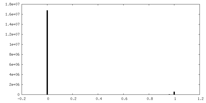
| Final reconstruction | Resolution.type: BY AUTHOR / Resolution: 3.7 Å / Resolution method: FSC 0.143 CUT-OFF / Number images used: 58448 |
|---|---|
| Initial angle assignment | Type: MAXIMUM LIKELIHOOD |
| Final angle assignment | Type: MAXIMUM LIKELIHOOD |
| FSC plot (resolution estimation) | 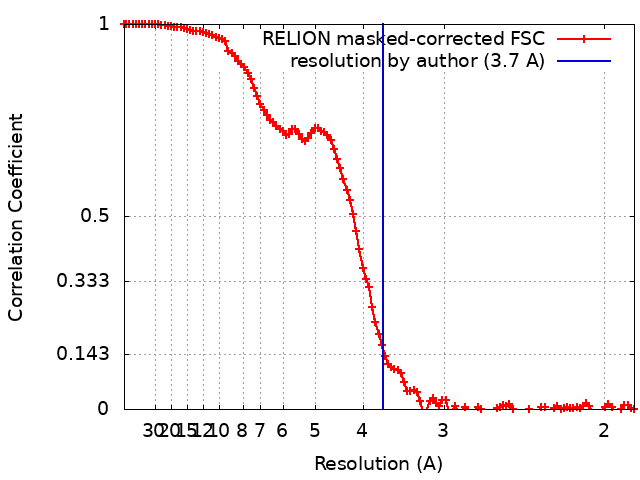 |
