 Movie
Movie Controller

[English] 日本語
 Yorodumi
- EMDB-21692: PKA RIIbeta holoenzyme with DnaJB1-PKAc fusion in fibrolamellar h... -
+ Open data

- Basic information
| Entry | Database: EMDB / ID: EMD-21692 | ||||||||||||
|---|---|---|---|---|---|---|---|---|---|---|---|---|---|
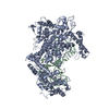
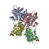
| Title | PKA RIIbeta holoenzyme with DnaJB1-PKAc fusion in fibrolamellar hepatoceullar carcinoma | ||||||||||||
 Map data Map data | C1-symmetry map | ||||||||||||
 Sample Sample |
| ||||||||||||
 Keywords Keywords | fibrolamellar hepatoceullar carcinoma / PKA / cAMP / Kinase / SIGNALING PROTEIN | ||||||||||||
| Function / homology |  Function and homology information Function and homology informationPKA activation in glucagon signalling / CREB1 phosphorylation through the activation of Adenylate Cyclase / DARPP-32 events / GPER1 signaling / Loss of Nlp from mitotic centrosomes / Recruitment of mitotic centrosome proteins and complexes / Loss of proteins required for interphase microtubule organization from the centrosome / Recruitment of NuMA to mitotic centrosomes / Anchoring of the basal body to the plasma membrane / AURKA Activation by TPX2 ...PKA activation in glucagon signalling / CREB1 phosphorylation through the activation of Adenylate Cyclase / DARPP-32 events / GPER1 signaling / Loss of Nlp from mitotic centrosomes / Recruitment of mitotic centrosome proteins and complexes / Loss of proteins required for interphase microtubule organization from the centrosome / Recruitment of NuMA to mitotic centrosomes / Anchoring of the basal body to the plasma membrane / AURKA Activation by TPX2 / Factors involved in megakaryocyte development and platelet production / Regulation of PLK1 Activity at G2/M Transition / Hedgehog 'off' state / PKA activation / positive regulation of protein folding / PKA-mediated phosphorylation of CREB / PKA-mediated phosphorylation of key metabolic factors / ROBO receptors bind AKAP5 / sperm head / negative regulation of inclusion body assembly / HDL assembly / channel activator activity / cAMP-dependent protein kinase regulator activity / mitochondrial protein catabolic process / Regulation of glycolysis by fructose 2,6-bisphosphate metabolism / response to antipsychotic drug / cell communication by electrical coupling involved in cardiac conduction / high-density lipoprotein particle assembly / nucleotide-activated protein kinase complex / Rap1 signalling / potassium channel inhibitor activity / High laminar flow shear stress activates signaling by PIEZO1 and PECAM1:CDH5:KDR in endothelial cells / Vasopressin regulates renal water homeostasis via Aquaporins / cAMP-dependent protein kinase inhibitor activity / histone H1-4S35 kinase activity / cAMP-dependent protein kinase / regulation of protein processing / cAMP-dependent protein kinase activity / cellular response to parathyroid hormone stimulus / protein localization to lipid droplet / negative regulation of interleukin-2 production / regulation of bicellular tight junction assembly / cAMP-dependent protein kinase complex / dorsal/ventral neural tube patterning / Loss of phosphorylation of MECP2 at T308 / CREB1 phosphorylation through the activation of Adenylate Cyclase / PKA activation / Triglyceride catabolism / regulation of osteoblast differentiation / sperm capacitation / cellular response to cold / ciliary base / protein kinase A regulatory subunit binding / negative regulation of glycolytic process through fructose-6-phosphate / protein kinase A catalytic subunit binding / forebrain development / ATPase activator activity / intracellular potassium ion homeostasis / mesoderm formation / RET signaling / Interleukin-3, Interleukin-5 and GM-CSF signaling / PKA activation in glucagon signalling / HSF1-dependent transactivation / regulation of cardiac conduction / plasma membrane raft / Regulation of HSF1-mediated heat shock response / response to unfolded protein / Regulation of MECP2 expression and activity / DARPP-32 events / regulation of macroautophagy / Attenuation phase / cAMP/PKA signal transduction / regulation of cardiac muscle contraction / sperm flagellum / postsynaptic modulation of chemical synaptic transmission / cAMP binding / transcription regulator inhibitor activity / vascular endothelial cell response to laminar fluid shear stress / renal water homeostasis / negative regulation of cAMP/PKA signal transduction / Hedgehog 'off' state / regulation of cellular response to heat / Ion homeostasis / negative regulation of protein localization to chromatin / negative regulation of TORC1 signaling / regulation of cardiac muscle contraction by regulation of the release of sequestered calcium ion / protein serine/threonine/tyrosine kinase activity / Loss of Nlp from mitotic centrosomes / Loss of proteins required for interphase microtubule organization from the centrosome / regulation of proteasomal protein catabolic process / Recruitment of mitotic centrosome proteins and complexes / protein export from nucleus / cellular response to epinephrine stimulus / protein folding chaperone / cellular response to glucagon stimulus / Recruitment of NuMA to mitotic centrosomes / calcium channel complex / CD209 (DC-SIGN) signaling / Anchoring of the basal body to the plasma membrane / Mitochondrial protein degradation Similarity search - Function | ||||||||||||
| Biological species |  Homo sapiens (human) / Homo sapiens (human) /  | ||||||||||||
| Method | single particle reconstruction / cryo EM / Resolution: 7.5 Å | ||||||||||||
 Authors Authors | Lu T-W / Aoto PC | ||||||||||||
| Funding support |  United States, 3 items United States, 3 items
| ||||||||||||
 Citation Citation | Journal: PLoS Biol / Year: 2020 Title: Structural analyses of the PKA RIIβ holoenzyme containing the oncogenic DnaJB1-PKAc fusion protein reveal protomer asymmetry and fusion-induced allosteric perturbations in fibrolamellar hepatocellular carcinoma. Authors: Tsan-Wen Lu / Phillip C Aoto / Jui-Hung Weng / Cole Nielsen / Jennifer N Cash / James Hall / Ping Zhang / Sanford M Simon / Michael A Cianfrocco / Susan S Taylor / Abstract: When the J-domain of the heat shock protein DnaJB1 is fused to the catalytic (C) subunit of cAMP-dependent protein kinase (PKA), replacing exon 1, this fusion protein, J-C subunit (J-C), becomes the ...When the J-domain of the heat shock protein DnaJB1 is fused to the catalytic (C) subunit of cAMP-dependent protein kinase (PKA), replacing exon 1, this fusion protein, J-C subunit (J-C), becomes the driver of fibrolamellar hepatocellular carcinoma (FL-HCC). Here, we use cryo-electron microscopy (cryo-EM) to characterize J-C bound to RIIβ, the major PKA regulatory (R) subunit in liver, thus reporting the first cryo-EM structure of any PKA holoenzyme. We report several differences in both structure and dynamics that could not be captured by the conventional crystallography approaches used to obtain prior structures. Most striking is the asymmetry caused by the absence of the second cyclic nucleotide binding (CNB) domain and the J-domain in one of the RIIβ:J-C protomers. Using molecular dynamics (MD) simulations, we discovered that this asymmetry is already present in the wild-type (WT) RIIβ2C2 but had been masked in the previous crystal structure. This asymmetry may link to the intrinsic allosteric regulation of all PKA holoenzymes and could also explain why most disease mutations in PKA regulatory subunits are dominant negative. The cryo-EM structure, combined with small-angle X-ray scattering (SAXS), also allowed us to predict the general position of the Dimerization/Docking (D/D) domain, which is essential for localization and interacting with membrane-anchored A-Kinase-Anchoring Proteins (AKAPs). This position provides a multivalent mechanism for interaction of the RIIβ holoenzyme with membranes and would be perturbed in the oncogenic fusion protein. The J-domain also alters several biochemical properties of the RIIβ holoenzyme: It is easier to activate with cAMP, and the cooperativity is reduced. These results provide new insights into how the finely tuned allosteric PKA signaling network is disrupted by the oncogenic J-C subunit, ultimately leading to the development of FL-HCC. | ||||||||||||
| History |
|
- Structure visualization
| Movie |
Movie viewer |
|---|---|
| Structure viewer | EM map: SurfViewMolmilJmol/JSmol |
| Supplemental images |

- Downloads & links
-EMDB archive
| Map data | emd_21692.map.gz | 40.3 MB | EMDB map data format | |
|---|---|---|---|---|
| Header (meta data) | emd-21692-v30.xmlemd-21692.xml | 13.1 KB 13.1 KB | Display Display | EMDB header |
| Images | 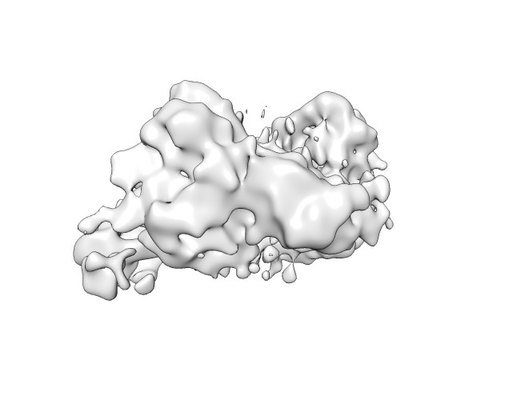 emd_21692.png emd_21692.png | 40.3 KB | ||
| Filedesc metadata | emd-21692.cif.gz | 5.8 KB | ||
| Archive directory |  http://ftp.pdbj.org/pub/emdb/structures/EMD-21692ftp://ftp.pdbj.org/pub/emdb/structures/EMD-21692 http://ftp.pdbj.org/pub/emdb/structures/EMD-21692ftp://ftp.pdbj.org/pub/emdb/structures/EMD-21692 | HTTPS FTP |
-Related structure data
| Related structure data |  6wjfMC  6wjgC M: atomic model generated by this map C: citing same article ( |
|---|---|
| Similar structure data |


-Links
| EMDB pages | EMDB (EBI/PDBe) / EMDataResource |
|---|---|
| Related items in Molecule of the Month |


-Map
| File | Download / File: emd_21692.map.gz / Format: CCP4 / Size: 64 MB / Type: IMAGE STORED AS FLOATING POINT NUMBER (4 BYTES) | ||||||||||||||||||||||||||||||||||||||||||||||||||||||||||||||||||||
|---|---|---|---|---|---|---|---|---|---|---|---|---|---|---|---|---|---|---|---|---|---|---|---|---|---|---|---|---|---|---|---|---|---|---|---|---|---|---|---|---|---|---|---|---|---|---|---|---|---|---|---|---|---|---|---|---|---|---|---|---|---|---|---|---|---|---|---|---|---|
| Annotation | C1-symmetry map | ||||||||||||||||||||||||||||||||||||||||||||||||||||||||||||||||||||
| Projections & slices | Image control
Images are generated by Spider. | ||||||||||||||||||||||||||||||||||||||||||||||||||||||||||||||||||||
| Voxel size | X=Y=Z: 1 Å | ||||||||||||||||||||||||||||||||||||||||||||||||||||||||||||||||||||
| Density |
| ||||||||||||||||||||||||||||||||||||||||||||||||||||||||||||||||||||
| Symmetry | Space group: 1 | ||||||||||||||||||||||||||||||||||||||||||||||||||||||||||||||||||||
| Details | EMDB XML:
CCP4 map header:
| ||||||||||||||||||||||||||||||||||||||||||||||||||||||||||||||||||||
 Z (Sec.)
 Y (Row.)
 X (Col.)


-Supplemental data
- Sample components
-Entire : PKA RIIbeta holoenzyme with DnaJB1-PKAc fusion in fibrolamellar h...
| Entire | Name: PKA RIIbeta holoenzyme with DnaJB1-PKAc fusion in fibrolamellar hepatoceullar carcinoma |
|---|---|
| Components |
|
-Supramolecule #1: PKA RIIbeta holoenzyme with DnaJB1-PKAc fusion in fibrolamellar h...
| Supramolecule | Name: PKA RIIbeta holoenzyme with DnaJB1-PKAc fusion in fibrolamellar hepatoceullar carcinoma type: complex / ID: 1 / Parent: 0 / Macromolecule list: all |
|---|
-Supramolecule #2: DnaJ homolog subfamily B member 1,cAMP-dependent protein kinase c...
| Supramolecule | Name: DnaJ homolog subfamily B member 1,cAMP-dependent protein kinase catalytic subunit alpha fusion type: complex / ID: 2 / Parent: 1 / Macromolecule list: #1 |
|---|---|
| Source (natural) | Organism: Homo sapiens (human) |
-Supramolecule #3: cAMP-dependent protein kinase type II-beta regulatory subunit
| Supramolecule | Name: cAMP-dependent protein kinase type II-beta regulatory subunit type: complex / ID: 3 / Parent: 1 / Macromolecule list: #2 |
|---|---|
| Source (natural) | Organism: |
-Macromolecule #1: DnaJ homolog subfamily B member 1,cAMP-dependent protein kinase c...
| Macromolecule | Name: DnaJ homolog subfamily B member 1,cAMP-dependent protein kinase catalytic subunit alpha fusion type: protein_or_peptide / ID: 1 / Number of copies: 2 / Enantiomer: LEVO / EC number: cAMP-dependent protein kinase |
|---|---|
| Source (natural) | Organism: Homo sapiens (human) |
| Molecular weight | Theoretical: 47.337984 KDa |
| Recombinant expression | Organism:  |
| Sequence | String: GKDYYQTLGL ARGASDEEIK RAYRRQALRY HPDKNKEPGA EEKFKEIAEA YDVLSDPRKR EIFDRYGEEV KEFLAKAKED FLKKWESPA QNTAHLDQFE RIKTLGTGSF GRVMLVKHKE TGNHYAMKIL DKQKVVKLKQ IEHTLNEKRI LQAVNFPFLV K LEFSFKDN ...String: GKDYYQTLGL ARGASDEEIK RAYRRQALRY HPDKNKEPGA EEKFKEIAEA YDVLSDPRKR EIFDRYGEEV KEFLAKAKED FLKKWESPA QNTAHLDQFE RIKTLGTGSF GRVMLVKHKE TGNHYAMKIL DKQKVVKLKQ IEHTLNEKRI LQAVNFPFLV K LEFSFKDN SNLYMVMEYV PGGEMFSHLR RIGRFSEPHA RFYAAQIVLT FEYLHSLDLI YRDLKPENLL IDQQGYIQVT DF GFAKRVK GRTWTLCGTP EYLAPEIILS KGYNKGVDWW ALGVLIYEMA AGYPPFFADQ PIQIYEKIVS GKVRFPSHFS SDL KDLLRN LLQVDLTKRF GNLKNGVNDI KNHKWFATTD WIAIYQRKVE APFIPKFKGP GDTSNFDDYE EEEIRVSINE KCGK EFSEF UniProtKB: DnaJ homolog subfamily B member 1, cAMP-dependent protein kinase catalytic subunit alpha |
-Macromolecule #2: cAMP-dependent protein kinase type II-beta regulatory subunit
| Macromolecule | Name: cAMP-dependent protein kinase type II-beta regulatory subunit type: protein_or_peptide / ID: 2 / Number of copies: 2 / Enantiomer: LEVO |
|---|---|
| Source (natural) | Organism: |
| Molecular weight | Theoretical: 46.177852 KDa |
| Recombinant expression | Organism: |
| Sequence | String: MSIEIPAGLT ELLQGFTVEV LRHQPADLLE FALQHFTRLQ QENERKGAAR FGHEGRTWGD AGAAAGGGTP SKGVNFAEEP MRSDSENGE EEEAAEAGAF NAPVINRFTR RASVCAEAYN PDEEEDDAES RIIHPKTDDQ RNRLQEACKD ILLFKNLDPE Q MSQVLDAM ...String: MSIEIPAGLT ELLQGFTVEV LRHQPADLLE FALQHFTRLQ QENERKGAAR FGHEGRTWGD AGAAAGGGTP SKGVNFAEEP MRSDSENGE EEEAAEAGAF NAPVINRFTR RASVCAEAYN PDEEEDDAES RIIHPKTDDQ RNRLQEACKD ILLFKNLDPE Q MSQVLDAM FEKLVKEGEH VIDQGDDGDN FYVIDRGTFD IYVKCDGVGR CVGNYDNRGS FGELALMYNT PRAATITATS PG ALWGLDR VTFRRIIVKN NAKKRKMYES FIESLPFLKS LEVSERLKVV DVIGTKVYND GEQIIAQGDS ADSFFIVESG EVR ITMKRK GKSDIEENGA VEIARCLRGQ YFGELALVTN KPRAASAHAI GTVKCLAMDV QAFERLLGPC MEIMKRNIAT YEEQ LVALF GTNMDIVEPT A UniProtKB: cAMP-dependent protein kinase type II-beta regulatory subunit |
-Experimental details
-Structure determination
| Method | cryo EM |
|---|---|
 Processing Processing | single particle reconstruction |
| Aggregation state | particle |
-Sample preparation
| Buffer | pH: 5.8 |
|---|---|
| Vitrification | Cryogen name: ETHANE |
- Electron microscopy
| Microscope | FEI TITAN KRIOS |
|---|---|
| Image recording | Film or detector model: GATAN K2 SUMMIT (4k x 4k) / Detector mode: COUNTING / Average electron dose: 80.0 e/Å2 |
| Electron beam | Acceleration voltage: 300 kV / Electron source:  FIELD EMISSION GUN FIELD EMISSION GUN |
| Electron optics | Illumination mode: FLOOD BEAM / Imaging mode: BRIGHT FIELD |
| Experimental equipment |  Model: Titan Krios / Image courtesy: FEI Company |
-Image processing
| Startup model | Type of model: OTHER / Details: Stochastic gradient descent |
|---|---|
| Final reconstruction | Applied symmetry - Point group: C1 (asymmetric) / Resolution.type: BY AUTHOR / Resolution: 7.5 Å / Resolution method: FSC 0.143 CUT-OFF / Number images used: 11182 |
| Initial angle assignment | Type: MAXIMUM LIKELIHOOD |
| Final angle assignment | Type: MAXIMUM LIKELIHOOD |
