National Institutes of Health/National Institute of General Medical Sciences (NIH/NIGMS)
GM103368
United States
Leona M. and Harry B. Helmsley Charitble Trust Grant
#2012-PG-MED002
United States
National Institutes of Health/National Institute of Diabetes and Digestive and Kidney Disease (NIH/NIDDK)
Intramural Program of the National Institute of Diabetes and Digestive Diseases
United States
Citation
Journal: Science / Year: 2017 Title: Cryo-EM structures and atomic model of the HIV-1 strand transfer complex intasome. Authors: Dario Oliveira Passos / Min Li / Renbin Yang / Stephanie V Rebensburg / Rodolfo Ghirlando / Youngmin Jeon / Nikoloz Shkriabai / Mamuka Kvaratskhelia / Robert Craigie / Dmitry Lyumkis / Abstract: Like all retroviruses, HIV-1 irreversibly inserts a viral DNA (vDNA) copy of its RNA genome into host target DNA (tDNA). The intasome, a higher-order nucleoprotein complex composed of viral integrase ...Like all retroviruses, HIV-1 irreversibly inserts a viral DNA (vDNA) copy of its RNA genome into host target DNA (tDNA). The intasome, a higher-order nucleoprotein complex composed of viral integrase (IN) and the ends of linear vDNA, mediates integration. Productive integration into host chromatin results in the formation of the strand transfer complex (STC) containing catalytically joined vDNA and tDNA. HIV-1 intasomes have been refractory to high-resolution structural studies. We used a soluble IN fusion protein to facilitate structural studies, through which we present a high-resolution cryo-electron microscopy (cryo-EM) structure of the core tetrameric HIV-1 STC and a higher-order form that adopts carboxyl-terminal domain rearrangements. The distinct STC structures highlight how HIV-1 can use the common retroviral intasome core architecture to accommodate different IN domain modules for assembly.
History
Deposition
Nov 28, 2016
-
Header (metadata) release
Jan 11, 2017
-
Map release
Jan 11, 2017
-
Update
Mar 13, 2024
-
Current status
Mar 13, 2024
Processing site: RCSB / Status: Released
-
Structure visualization
Movie
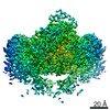
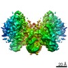
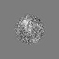
Surface view with section colored by density value
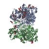
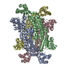
Entire : complex formed by a tetrameric assembly of Sso7d-fusion HIV-1 Int...
Entire
Name: complex formed by a tetrameric assembly of Sso7d-fusion HIV-1 Integrase with the product of DNA strand transfer
Components
Complex: complex formed by a tetrameric assembly of Sso7d-fusion HIV-1 Integrase with the product of DNA strand transfer
Protein or peptide: HIV-1 Integrase, Sso7d chimera
DNA: DNA (11-MER)
DNA: DNA (23-MER)
DNA: DNA (37-MER)
Ligand: ZINC ION
Ligand: MAGNESIUM ION
-
Supramolecule #1: complex formed by a tetrameric assembly of Sso7d-fusion HIV-1 Int...
Supramolecule
Name: complex formed by a tetrameric assembly of Sso7d-fusion HIV-1 Integrase with the product of DNA strand transfer type: complex / ID: 1 / Parent: 0 / Macromolecule list: #1-#4
Source (natural)
Organism: Human immunodeficiency virus 1
Molecular weight
Theoretical: 228 KDa
-
Macromolecule #1: HIV-1 Integrase, Sso7d chimera
Macromolecule
Name: HIV-1 Integrase, Sso7d chimera / type: protein_or_peptide / ID: 1 / Number of copies: 4 / Enantiomer: LEVO
Cryogen name: ETHANE / Chamber humidity: 50 % / Chamber temperature: 277 K / Instrument: HOMEMADE PLUNGER Details: Sample containing HIV STC intasomes in SEC buffer was applied onto freshly plasma-treated (6 seconds, Gatan Solarus plasma cleaner) holey gold UltrAuFoil grids (Quantifoil), adsorbed for 30 ...Details: Sample containing HIV STC intasomes in SEC buffer was applied onto freshly plasma-treated (6 seconds, Gatan Solarus plasma cleaner) holey gold UltrAuFoil grids (Quantifoil), adsorbed for 30 seconds, then plunged into liquid ethane using a manual cryo-plunger in an ambient environment of 4 degrees C..
-
Electron microscopy
Microscope
FEI TITAN KRIOS
Temperature
Min: 90.0 K / Max: 90.0 K
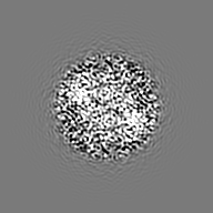
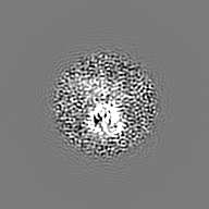
Image recording
Film or detector model: GATAN K2 SUMMIT (4k x 4k) / Detector mode: COUNTING / Digitization - Dimensions - Width: 3838 pixel / Digitization - Dimensions - Height: 3710 pixel / Digitization - Frames/image: 1-100 / Number grids imaged: 1 / Number real images: 1225 / Average exposure time: 20.0 sec. / Average electron dose: 95.0 e/Å2 Details: Individual frames were gain-corrected, aligned, and summed with the application of an exposure filter using MotionCor2, according to the nominal dose rate.
Electron beam
Acceleration voltage: 300 kV / Electron source: FIELD EMISSION GUN
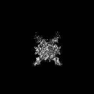
Type of model: INSILICO MODEL / In silico model: common lines model using OptiMod Details: An initial model was generated directly from the class averages using OptiMod.
Final reconstruction
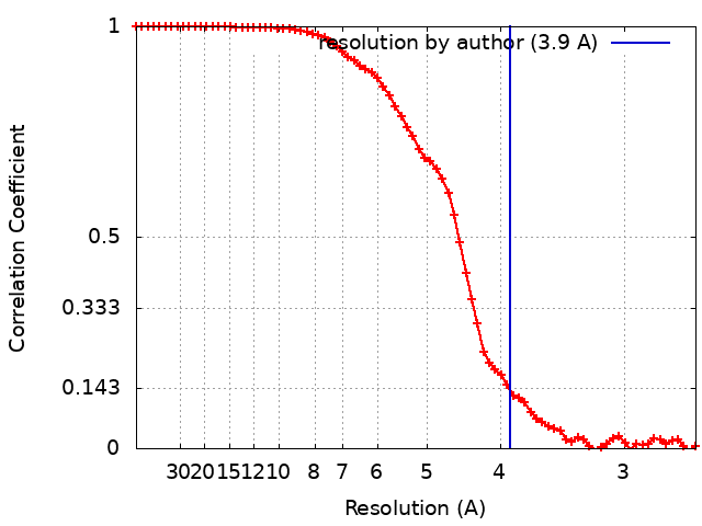
Applied symmetry - Point group: C2 (2 fold cyclic) / Algorithm: FOURIER SPACE / Resolution.type: BY AUTHOR / Resolution: 3.9 Å / Resolution method: FSC 0.143 CUT-OFF / Software - Name: FREALIGN (ver. 9.11) / Details: Resolution-limited refinement used throughout / Number images used: 83766
Type: PROJECTION MATCHING / Software - Name: FREALIGN (ver. 9.11) / Details: Frealign 3D classification and refinement
Final 3D classification
Software - Name: FREALIGN (ver. 3.11)
FSC plot (resolution estimation)
-
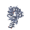
Atomic model buiding 1
Details
To generate ensemble models, the complete intasome model was iteratively relaxed - using two-fold symmetry and a combination of Rosetta and Phenix - against one half map (the working map) and inspected for consistency with the second half map (the free map). The model was then adjusted manually using Coot. Final ensemble modeling used half maps for all aspects of refinement and evaluation: 500 models were generated as described using Rosetta. From the 100 top-scoring models (scored by Rosetta energy), the ten models with the best map-to-model FSC were selected and refined in real space using secondary-structure restraints in Phenix. Molprobity was used throughout the refinement process.
Refinement
Space: REAL / Protocol: FLEXIBLE FIT / Overall B value: 180 / Target criteria: FSC 0.5
Output model
PDB-5u1c: Structure of tetrameric HIV-1 Strand Transfer Complex Intasome
+
About Yorodumi
-
News
-
Feb 9, 2022. New format data for meta-information of EMDB entries
New format data for meta-information of EMDB entries
Version 3 of the EMDB header file is now the official format.
The previous official version 1.9 will be removed from the archive.
In the structure databanks used in Yorodumi, some data are registered as the other names, "COVID-19 virus" and "2019-nCoV". Here are the details of the virus and the list of structure data.
Jan 31, 2019. EMDB accession codes are about to change! (news from PDBe EMDB page)
EMDB accession codes are about to change! (news from PDBe EMDB page)
The allocation of 4 digits for EMDB accession codes will soon come to an end. Whilst these codes will remain in use, new EMDB accession codes will include an additional digit and will expand incrementally as the available range of codes is exhausted. The current 4-digit format prefixed with “EMD-” (i.e. EMD-XXXX) will advance to a 5-digit format (i.e. EMD-XXXXX), and so on. It is currently estimated that the 4-digit codes will be depleted around Spring 2019, at which point the 5-digit format will come into force.
The EM Navigator/Yorodumi systems omit the EMD- prefix.
Related info.:Q: What is EMD? / ID/Accession-code notation in Yorodumi/EM Navigator
Yorodumi is a browser for structure data from EMDB, PDB, SASBDB, etc.
This page is also the successor to EM Navigator detail page, and also detail information page/front-end page for Omokage search.
The word "yorodu" (or yorozu) is an old Japanese word meaning "ten thousand". "mi" (miru) is to see.
Related info.:EMDB / PDB / SASBDB / Comparison of 3 databanks / Yorodumi Search / Aug 31, 2016. New EM Navigator & Yorodumi / Yorodumi Papers / Jmol/JSmol / Function and homology information / Changes in new EM Navigator and Yorodumi
 Movie
Movie Controller


 Open data
 Basic information
 Map data
 Sample
 Keywords
 Function and homology information
 Human immunodeficiency virus 1 /
  Homo sapiens (human)
 Authors
 United States, 3 items
  Citation
 Structure visualization
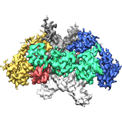
 Downloads & links
 emd_8481.png
 http://ftp.pdbj.org/pub/emdb/structures/EMD-8481


 Z (Sec.)
 Y (Row.)
 X (Col.)


 Sample components
 Processing
 Electron microscopy
 FIELD EMISSION GUN