 Movie
Movie Controller


+ Open data

- Basic information
| Entry | Database: PDB / ID: 7ppo | ||||||
|---|---|---|---|---|---|---|---|
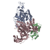
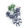
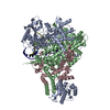
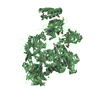
| Title | Structure of SidJ/CaM bound to SdeA in pre-glutamylation state | ||||||
 Components Components |
| ||||||
 Keywords Keywords | LIGASE / Glutamylase / Pseudokinase / phosphoribosyl / ubiquitination / Complex | ||||||
| Function / homology |  Function and homology information Function and homology informationprotein-glutamic acid ligase activity, initiating / protein-glutamic acid ligase activity, elongating / Ligases / NAD+-protein-arginine ADP-ribosyltransferase / transporter inhibitor activity / NAD+-protein-arginine ADP-ribosyltransferase activity / deNEDDylase activity / protein deneddylation / Transferases; Acyltransferases; Aminoacyltransferases / K63-linked deubiquitinase activity ...protein-glutamic acid ligase activity, initiating / protein-glutamic acid ligase activity, elongating / Ligases / NAD+-protein-arginine ADP-ribosyltransferase / transporter inhibitor activity / NAD+-protein-arginine ADP-ribosyltransferase activity / deNEDDylase activity / protein deneddylation / Transferases; Acyltransferases; Aminoacyltransferases / K63-linked deubiquitinase activity / negative regulation of ryanodine-sensitive calcium-release channel activity / negative regulation of calcium ion export across plasma membrane / presynaptic endocytosis / regulation of cell communication by electrical coupling involved in cardiac conduction / calcineurin-mediated signaling / adenylate cyclase binding / protein phosphatase activator activity / protein deubiquitination / detection of calcium ion / regulation of cardiac muscle contraction / catalytic complex / regulation of cardiac muscle contraction by regulation of the release of sequestered calcium ion / calcium channel inhibitor activity / presynaptic cytosol / regulation of release of sequestered calcium ion into cytosol by sarcoplasmic reticulum / titin binding / regulation of calcium-mediated signaling / sperm midpiece / voltage-gated potassium channel complex / nucleotidyltransferase activity / cysteine-type peptidase activity / calcium channel complex / substantia nigra development / regulation of heart rate / calyx of Held / adenylate cyclase activator activity / sarcomere / protein serine/threonine kinase activator activity / regulation of cytokinesis / spindle microtubule / calcium channel regulator activity / response to calcium ion / G2/M transition of mitotic cell cycle / long-term synaptic potentiation / spindle pole / calcium-dependent protein binding / myelin sheath / host cell / transferase activity / vesicle / transmembrane transporter binding / Hydrolases; Acting on peptide bonds (peptidases); Cysteine endopeptidases / protein ubiquitination / G protein-coupled receptor signaling pathway / nucleotide binding / calcium ion binding / centrosome / protein kinase binding / protein-containing complex / proteolysis / extracellular region / metal ion binding / nucleus / membrane / plasma membrane / cytoplasm Similarity search - Function | ||||||
| Biological species |   Legionella pneumophila (bacteria) Legionella pneumophila (bacteria) Homo sapiens (human) Homo sapiens (human) | ||||||
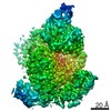
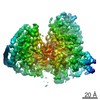
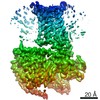
| Method | ELECTRON MICROSCOPY / single particle reconstruction / cryo EM / Resolution: 2.91 Å | ||||||
 Authors Authors | Adams, M. / Bhogaraju, S. | ||||||
| Funding support | European Union, 1items
| ||||||
 Citation Citation | Journal: Nat Commun / Year: 2021 Title: Structural basis for protein glutamylation by the Legionella pseudokinase SidJ. Authors: Michael Adams / Rahul Sharma / Thomas Colby / Felix Weis / Ivan Matic / Sagar Bhogaraju /   Abstract: Legionella pneumophila (LP) avoids phagocytosis by secreting nearly 300 effector proteins into the host cytosol. SidE family of effectors (SdeA, SdeB, SdeC and SidE) employ phosphoribosyl ...Legionella pneumophila (LP) avoids phagocytosis by secreting nearly 300 effector proteins into the host cytosol. SidE family of effectors (SdeA, SdeB, SdeC and SidE) employ phosphoribosyl ubiquitination to target multiple host Rab GTPases and innate immune factors. To suppress the deleterious toxicity of SidE enzymes in a timely manner, LP employs a metaeffector named SidJ. Upon activation by host Calmodulin (CaM), SidJ executes an ATP-dependent glutamylation to modify the catalytic residue Glu860 in the mono-ADP-ribosyl transferase (mART) domain of SdeA. SidJ is a unique glutamylase that adopts a kinase-like fold but contains two nucleotide-binding pockets. There is a lack of consensus about the substrate recognition and catalytic mechanism of SidJ. Here, we determined the cryo-EM structure of SidJ in complex with its substrate SdeA in two different states of catalysis. Our structures reveal that both phosphodiesterase (PDE) and mART domains of SdeA make extensive contacts with SidJ. In the pre-glutamylation state structure of the SidJ-SdeA complex, adenylylated E860 of SdeA is inserted into the non-canonical (migrated) nucleotide-binding pocket of SidJ. Structure-based mutational analysis indicates that SidJ employs its migrated pocket for the glutamylation of SdeA. Finally, using mass spectrometry, we identified several transient autoAMPylation sites close to both the catalytic pockets of SidJ. Our data provide unique insights into the substrate recognition and the mechanism of protein glutamylation by the pseudokinase SidJ. | ||||||
| History |
|
- Structure visualization
| Movie |
Movie viewer |
|---|---|
| Structure viewer | Molecule: MolmilJmol/JSmol |
- Downloads & links
-Download
| PDBx/mmCIF format | 7ppo.cif.gz | 270.1 KB | Display | PDBx/mmCIF format |
|---|---|---|---|---|
| PDB format | pdb7ppo.ent.gz | 206.5 KB | Display | PDB format |
| PDBx/mmJSON format | 7ppo.json.gz | Tree view | PDBx/mmJSON format | |
| Others |  Other downloads Other downloads |
-Validation report
| Arichive directory | https://data.pdbj.org/pub/pdb/validation_reports/pp/7ppoftp://data.pdbj.org/pub/pdb/validation_reports/pp/7ppo | HTTPS FTP |
|---|
-Related structure data
| Related structure data |  13583MC 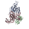 7pqeC M: map data used to model this data C: citing same article ( |
|---|---|
| Similar structure data |


-Links
 PDBj
PDBj- Assembly
| Deposited unit | 
|
|---|---|
| 1 |
|
-Components
| #1: Protein | Mass: 111200.375 Da / Num. of mol.: 1 Source method: isolated from a genetically manipulated source Source: (gene. exp.) Legionella pneumophila (bacteria) / Gene: sdeA, lpg2157 / Production host: References: UniProt: Q5ZTK4, Hydrolases; Acting on peptide bonds (peptidases); Cysteine endopeptidases, Transferases; Acyltransferases; Aminoacyltransferases, NAD+-protein-arginine ADP-ribosyltransferase |
|---|---|
| #2: Protein | Mass: 91826.992 Da / Num. of mol.: 1 / Mutation: E565A Source method: isolated from a genetically manipulated source Source: (gene. exp.) Legionella pneumophila (bacteria) / Gene: sidJ, lpg2155 / Production host: |
| #3: Protein | Mass: 19066.006 Da / Num. of mol.: 1 Source method: isolated from a genetically manipulated source Source: (gene. exp.) Homo sapiens (human) / Production host: |
| #4: Chemical | ChemComp-MG /   Mass: 24.305 Da / Num. of mol.: 1 / Source method: obtained synthetically / Formula: Mg Mass: 24.305 Da / Num. of mol.: 1 / Source method: obtained synthetically / Formula: Mg |
| #5: Chemical | ChemComp-CA /   Mass: 40.078 Da / Num. of mol.: 1 / Source method: obtained synthetically / Formula: Ca Mass: 40.078 Da / Num. of mol.: 1 / Source method: obtained synthetically / Formula: Ca |
| Has ligand of interest | Y |
-Experimental details
-Experiment
| Experiment | Method: ELECTRON MICROSCOPY |
|---|---|
| EM experiment | Aggregation state: PARTICLE / 3D reconstruction method: single particle reconstruction |
- Sample preparation
| Component | Name: SidJ/CaM-SdeA / Type: COMPLEX / Entity ID: #1-#3 / Source: RECOMBINANT |
|---|---|
| Source (natural) | Organism: Legionella pneumophila (bacteria) |
| Source (recombinant) | Organism: |
| Buffer solution | pH: 7.5 |
| Specimen | Embedding applied: NO / Shadowing applied: NO / Staining applied: NO / Vitrification applied: YES |
| Vitrification | Instrument: FEI VITROBOT MARK I / Cryogen name: ETHANE / Humidity: 100 % / Chamber temperature: 4 K |
- Electron microscopy imaging
| Experimental equipment |  Model: Titan Krios / Image courtesy: FEI Company |
|---|---|
| Microscopy | Model: FEI TITAN KRIOS |
| Electron gun | Electron source:  FIELD EMISSION GUN / Accelerating voltage: 300 kV / Illumination mode: FLOOD BEAM FIELD EMISSION GUN / Accelerating voltage: 300 kV / Illumination mode: FLOOD BEAM |
| Electron lens | Mode: BRIGHT FIELD |
| Image recording | Electron dose: 47.45 e/Å2 / Film or detector model: GATAN K3 (6k x 4k) |
- Processing
| Software |
| ||||||||||||||||||||||||
|---|---|---|---|---|---|---|---|---|---|---|---|---|---|---|---|---|---|---|---|---|---|---|---|---|---|
| CTF correction | Type: PHASE FLIPPING AND AMPLITUDE CORRECTION | ||||||||||||||||||||||||
| 3D reconstruction | Resolution: 2.91 Å / Resolution method: FSC 0.143 CUT-OFF / Num. of particles: 140022 / Symmetry type: POINT | ||||||||||||||||||||||||
| Refinement | Stereochemistry target values: GeoStd + Monomer Library + CDL v1.2 | ||||||||||||||||||||||||
| Refine LS restraints |
|
