Movie
Movie Controller


+ Open data

- Basic information
| Entry | Database: EMDB / ID: EMD-10933 | |||||||||
|---|---|---|---|---|---|---|---|---|---|---|
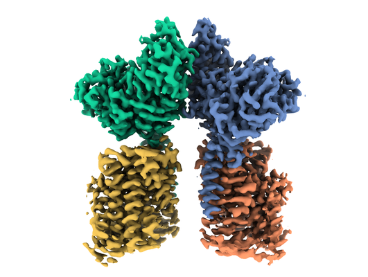
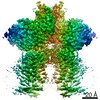
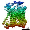
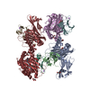
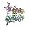
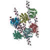
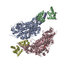
| Title | Heterotetrameric structure of the rBAT-b(0,+)AT1 complex | |||||||||
 Map data Map data | ||||||||||
 Sample Sample |
| |||||||||
 Keywords Keywords | Membrane transporter Heteromeric amino acid transporter Human transporter / MEMBRANE PROTEIN | |||||||||
| Function / homology |  Function and homology information Function and homology informationbroad specificity neutral L-amino acid:basic L-amino acid antiporter activity / Defective SLC3A1 causes cystinuria (CSNU) / Defective amino acid transport by SLC7A9 causes cystinuria (CSNU) / basic amino acid transport / L-cystine transmembrane transporter activity / basic amino acid transmembrane transporter activity / L-cystine transport / aspartate transmembrane transport / amino acid transmembrane transport / L-glutamate transmembrane transport ...broad specificity neutral L-amino acid:basic L-amino acid antiporter activity / Defective SLC3A1 causes cystinuria (CSNU) / Defective amino acid transport by SLC7A9 causes cystinuria (CSNU) / basic amino acid transport / L-cystine transmembrane transporter activity / basic amino acid transmembrane transporter activity / L-cystine transport / aspartate transmembrane transport / amino acid transmembrane transport / L-glutamate transmembrane transport / neutral amino acid transport / neutral L-amino acid transmembrane transporter activity / Amino acid transport across the plasma membrane / amino acid transmembrane transporter activity / antiporter activity / vacuolar membrane / Basigin interactions / amino acid transport / brush border membrane / peptide antigen binding / protein-containing complex assembly / carbohydrate metabolic process / gene expression / apical plasma membrane / protein heterodimerization activity / protein-containing complex binding / extracellular exosome / membrane / plasma membrane Similarity search - Function | |||||||||
| Biological species |  Homo sapiens (human) Homo sapiens (human) | |||||||||
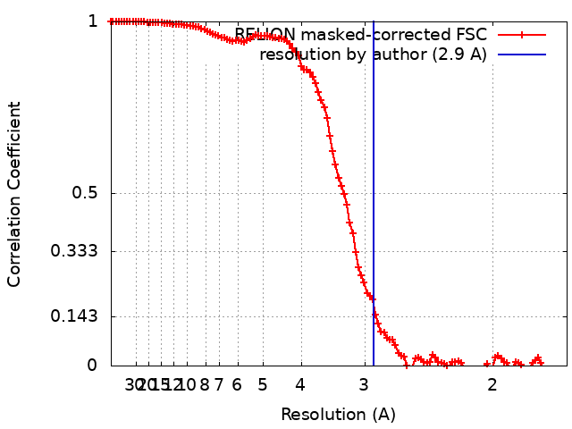
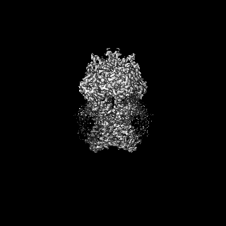
| Method | single particle reconstruction / cryo EM / Resolution: 2.9 Å | |||||||||
 Authors Authors | Wu D / Safarian S | |||||||||
| Funding support |  Germany, 2 items Germany, 2 items
| |||||||||
 Citation Citation | Journal: Proc Natl Acad Sci U S A / Year: 2020 Title: Structural basis for amino acid exchange by a human heteromeric amino acid transporter. Authors: Di Wu / Tamara N Grund / Sonja Welsch / Deryck J Mills / Max Michel / Schara Safarian / Hartmut Michel / Abstract: Heteromeric amino acid transporters (HATs) comprise a group of membrane proteins that belong to the solute carrier (SLC) superfamily. They are formed by two different protein components: a light ...Heteromeric amino acid transporters (HATs) comprise a group of membrane proteins that belong to the solute carrier (SLC) superfamily. They are formed by two different protein components: a light chain subunit from an SLC7 family member and a heavy chain subunit from the SLC3 family. The light chain constitutes the transport subunit whereas the heavy chain mediates trafficking to the plasma membrane and maturation of the functional complex. Mutation, malfunction, and dysregulation of HATs are associated with a wide range of pathologies or represent the direct cause of inherited and acquired disorders. Here we report the cryogenic electron microscopy structure of the neutral and basic amino acid transport complex (bAT1-rBAT) which reveals a heterotetrameric protein assembly composed of two heavy and light chain subunits, respectively. The previously uncharacterized interaction between two HAT units is mediated via dimerization of the heavy chain subunits and does not include participation of the light chain subunits. The bAT1 transporter adopts a LeuT fold and is captured in an inward-facing conformation. We identify an amino-acid-binding pocket that is formed by transmembrane helices 1, 6, and 10 and conserved among SLC7 transporters. | |||||||||
| History |
|
- Structure visualization
| Movie |
Movie viewer |
|---|---|
| Structure viewer | EM map: SurfViewMolmilJmol/JSmol |
| Supplemental images |
- Downloads & links
-EMDB archive
| Map data | emd_10933.map.gz | 113.2 MB | EMDB map data format | |
|---|---|---|---|---|
| Header (meta data) | emd-10933-v30.xmlemd-10933.xml | 18.4 KB 18.4 KB | Display Display | EMDB header |
| FSC (resolution estimation) | emd_10933_fsc.xml | 11.4 KB | Display | FSC data file |
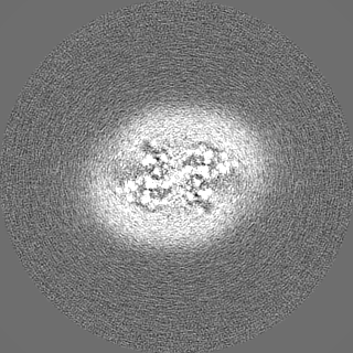
| Images |  emd_10933.png emd_10933.png | 172.7 KB | ||
| Filedesc metadata | emd-10933.cif.gz | 6.5 KB | ||
| Others | emd_10933_half_map_1.map.gzemd_10933_half_map_2.map.gz | 96.8 MB 96.8 MB | ||
| Archive directory |  http://ftp.pdbj.org/pub/emdb/structures/EMD-10933ftp://ftp.pdbj.org/pub/emdb/structures/EMD-10933 http://ftp.pdbj.org/pub/emdb/structures/EMD-10933ftp://ftp.pdbj.org/pub/emdb/structures/EMD-10933 | HTTPS FTP |
-Related structure data
| Related structure data |  6yupMC  6yuzC  6yv1C M: atomic model generated by this map C: citing same article ( |
|---|---|
| Similar structure data |


-Links
| EMDB pages | EMDB (EBI/PDBe) / EMDataResource |
|---|---|
| Related items in Molecule of the Month |


-Map
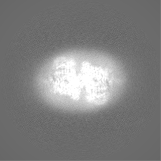
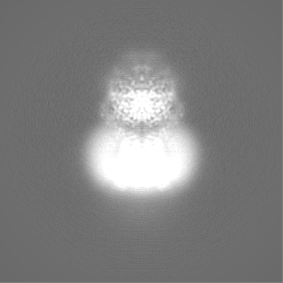
| File | Download / File: emd_10933.map.gz / Format: CCP4 / Size: 125 MB / Type: IMAGE STORED AS FLOATING POINT NUMBER (4 BYTES) | ||||||||||||||||||||||||||||||||||||||||||||||||||||||||||||
|---|---|---|---|---|---|---|---|---|---|---|---|---|---|---|---|---|---|---|---|---|---|---|---|---|---|---|---|---|---|---|---|---|---|---|---|---|---|---|---|---|---|---|---|---|---|---|---|---|---|---|---|---|---|---|---|---|---|---|---|---|---|
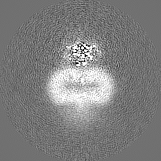
| Projections & slices | Image control
Images are generated by Spider. | ||||||||||||||||||||||||||||||||||||||||||||||||||||||||||||
| Voxel size | X=Y=Z: 0.837 Å | ||||||||||||||||||||||||||||||||||||||||||||||||||||||||||||
| Density |
| ||||||||||||||||||||||||||||||||||||||||||||||||||||||||||||
| Symmetry | Space group: 1 | ||||||||||||||||||||||||||||||||||||||||||||||||||||||||||||
| Details | EMDB XML:
CCP4 map header:
| ||||||||||||||||||||||||||||||||||||||||||||||||||||||||||||
 Z (Sec.)
 Y (Row.)
 X (Col.)


-Supplemental data
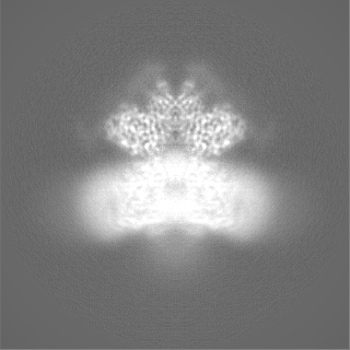
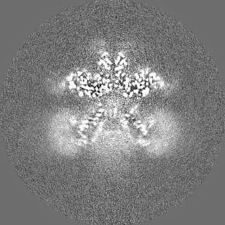
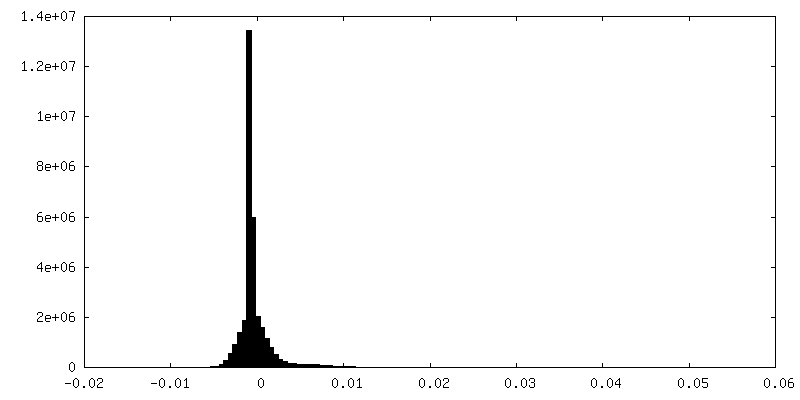
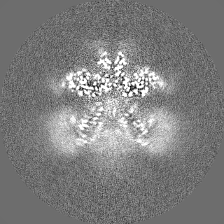
-Half map: #2
| File | emd_10933_half_map_1.map | ||||||||||||
|---|---|---|---|---|---|---|---|---|---|---|---|---|---|
| Projections & Slices |
| ||||||||||||
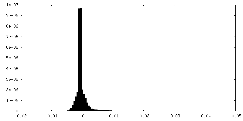
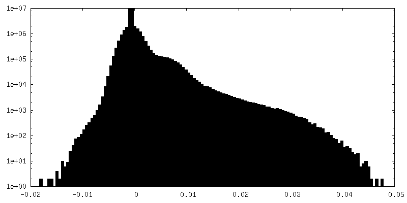
| Density Histograms |
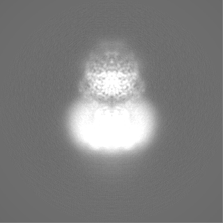
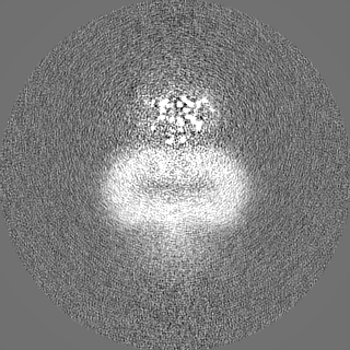
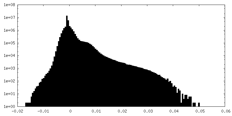
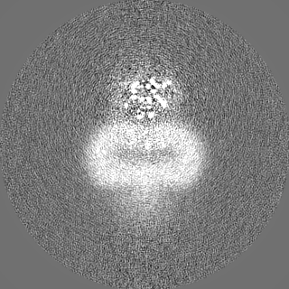
-Half map: #1
| File | emd_10933_half_map_2.map | ||||||||||||
|---|---|---|---|---|---|---|---|---|---|---|---|---|---|
| Projections & Slices |
| ||||||||||||
| Density Histograms |


- Sample components
-Entire : Heterotetrameric complex of rBAT and b(0,+)AT1
| Entire | Name: Heterotetrameric complex of rBAT and b(0,+)AT1 |
|---|---|
| Components |
|
-Supramolecule #1: Heterotetrameric complex of rBAT and b(0,+)AT1
| Supramolecule | Name: Heterotetrameric complex of rBAT and b(0,+)AT1 / type: complex / ID: 1 / Parent: 0 / Macromolecule list: #1-#2 / Details: 2 x rBAT 2 x b(0,+)AT1 |
|---|---|
| Source (natural) | Organism: Homo sapiens (human) |
| Molecular weight | Theoretical: 264.666 kDa/nm |
-Macromolecule #1: Neutral and basic amino acid transport protein rBAT
| Macromolecule | Name: Neutral and basic amino acid transport protein rBAT / type: protein_or_peptide / ID: 1 / Number of copies: 2 / Enantiomer: LEVO |
|---|---|
| Source (natural) | Organism: Homo sapiens (human) |
| Molecular weight | Theoretical: 78.937703 KDa |
| Recombinant expression | Organism: Homo sapiens (human) |
| Sequence | String: MAEDKSKRDS IEMSMKGCQT NNGFVHNEDI LEQTPDPGSS TDNLKHSTRG ILGSQEPDFK GVQPYAGMPK EVLFQFSGQA RYRIPREIL FWLTVASVLV LIAATIAIIA LSPKCLDWWQ EGPMYQIYPR SFKDSNKDGN GDLKGIQDKL DYITALNIKT V WITSFYKS ...String: MAEDKSKRDS IEMSMKGCQT NNGFVHNEDI LEQTPDPGSS TDNLKHSTRG ILGSQEPDFK GVQPYAGMPK EVLFQFSGQA RYRIPREIL FWLTVASVLV LIAATIAIIA LSPKCLDWWQ EGPMYQIYPR SFKDSNKDGN GDLKGIQDKL DYITALNIKT V WITSFYKS SLKDFRYGVE DFREVDPIFG TMEDFENLVA AIHDKGLKLI IDFIPNHTSD KHIWFQLSRT RTGKYTDYYI WH DCTHENG KTIPPNNWLS VYGNSSWHFD EVRNQCYFHQ FMKEQPDLNF RNPDVQEEIK EILRFWLTKG VDGFSLDAVK FLL EAKHLR DEIQVNKTQI PDTVTQYSEL YHDFTTTQVG MHDIVRSFRQ TMDQYSTEPG RYRFMGTEAY AESIDRTVMY YGLP FIQEA DFPFNNYLSM LDTVSGNSVY EVITSWMENM PEGKWPNWMI GGPDSSRLTS RLGNQYVNVM NMLLFTLPGT PITYY GEEI GMGNIVAANL NESYDINTLR SKSPMQWDNS SNAGFSEASN TWLPTNSDYH TVNVDVQKTQ PRSALKLYQD LSLLHA NEL LLNRGWFCHL RNDSHYVVYT RELDGIDRIF IVVLNFGEST LLNLHNMISG LPAKMRIRLS TNSADKGSKV DTSGIFL DK GEGLIFEHNT KNLLHRQTAF RDRCFVSNRA CYSSVLNILY TSC UniProtKB: Amino acid transporter heavy chain SLC3A1 |
-Macromolecule #2: b(0,+)-type amino acid transporter 1
| Macromolecule | Name: b(0,+)-type amino acid transporter 1 / type: protein_or_peptide / ID: 2 / Number of copies: 2 / Enantiomer: LEVO |
|---|---|
| Source (natural) | Organism: Homo sapiens (human) |
| Molecular weight | Theoretical: 53.515992 KDa |
| Recombinant expression | Organism: Homo sapiens (human) |
| Sequence | String: MGDTGLRKRR EDEKSIQSQE PKTTSLQKEL GLISGISIIV GTIIGSGIFV SPKSVLSNTE AVGPCLIIWA ACGVLATLGA LCFAELGTM ITKSGGEYPY LMEAYGPIPA YLFSWASLIV IKPTSFAIIC LSFSEYVCAP FYVGCKPPQI VVKCLAAAAI L FISTVNSL ...String: MGDTGLRKRR EDEKSIQSQE PKTTSLQKEL GLISGISIIV GTIIGSGIFV SPKSVLSNTE AVGPCLIIWA ACGVLATLGA LCFAELGTM ITKSGGEYPY LMEAYGPIPA YLFSWASLIV IKPTSFAIIC LSFSEYVCAP FYVGCKPPQI VVKCLAAAAI L FISTVNSL SVRLGSYVQN IFTAAKLVIV AIIIISGLVL LAQGNTKNFD NSFEGAQLSV GAISLAFYNG LWAYDGWNQL NY ITEELRN PYRNLPLAII IGIPLVTACY ILMNVSYFTV MTATELLQSQ AVAVTFGDRV LYPASWIVPL FVAFSTIGAA NGT CFTAGR LIYVAGREGH MLKVLSYISV RRLTPAPAII FYGIIATIYI IPGDINSLVN YFSFAAWLFY GLTILGLIVM RFTR KELER PIKVPVVIPV LMTLISVFLV LAPIISKPTW EYLYCVLFIL SGLLFYFLFV HYKFGWAQKI SKPITMHLQM LMEVV PPEE DPE UniProtKB: b(0,+)-type amino acid transporter 1 |
-Macromolecule #3: 2-acetamido-2-deoxy-beta-D-glucopyranose
| Macromolecule | Name: 2-acetamido-2-deoxy-beta-D-glucopyranose / type: ligand / ID: 3 / Number of copies: 8 / Formula: NAG |
|---|---|
| Molecular weight | Theoretical: 221.208 Da |
| Chemical component information |  ChemComp-NAG: |
-Macromolecule #4: CALCIUM ION
| Macromolecule | Name: CALCIUM ION / type: ligand / ID: 4 / Number of copies: 2 / Formula: CA |
|---|---|
| Molecular weight | Theoretical: 40.078 Da |
-Experimental details
-Structure determination
| Method | cryo EM |
|---|---|
 Processing Processing | single particle reconstruction |
| Aggregation state | particle |
-Sample preparation
| Concentration | 2.5 mg/mL | ||||||||
|---|---|---|---|---|---|---|---|---|---|
| Buffer | pH: 7.5 Component:
| ||||||||
| Grid | Model: C-flat-1.2/1.3 4C / Material: COPPER / Mesh: 400 / Support film - Material: CARBON / Support film - topology: HOLEY / Pretreatment - Type: GLOW DISCHARGE / Pretreatment - Time: 60 sec. | ||||||||
| Vitrification | Cryogen name: ETHANE / Chamber humidity: 100 % / Chamber temperature: 283.15 K / Instrument: FEI VITROBOT MARK IV |
- Electron microscopy
| Microscope | FEI TITAN KRIOS |
|---|---|
| Image recording | Film or detector model: GATAN K3 (6k x 4k) / Average electron dose: 40.0 e/Å2 |
| Electron beam | Acceleration voltage: 300 kV / Electron source:  FIELD EMISSION GUN FIELD EMISSION GUN |
| Electron optics | Illumination mode: FLOOD BEAM / Imaging mode: BRIGHT FIELD |
| Experimental equipment |  Model: Titan Krios / Image courtesy: FEI Company |