[English] 日本語
Yorodumi Papers
- Database of articles cited by EMDB/PDB/SASBDB data -

+
Search query

Keywords
Structure methods
Author
Journal
IF

-
Structure paper

TitleLarge-scale structural analysis of the classical human protein tyrosine phosphatome.
Journal, issue, pagesCell(Cambridge,Mass. ), Vol. 136, Page 352-363, Year 2009
Publish dateJul 28, 2005 (structure data deposition date)
AuthorsBarr, A.J. / Ugochukwu, E. / Lee, W.H. / King, O.N. / Filippakopoulos, P. / Alfano, I. / Savitsky, P. / Burgess-Brown, N.A. / Muller, S. / Knapp, S.
External linksCell(Cambridge,Mass. ) / PubMed:19167335
MethodsX-ray diffraction
Resolution1.5 - 3.2 Å
Structure data

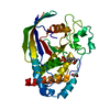
PDB-2ahs:
Crystal Structure of the Catalytic Domain of Human Tyrosine Receptor Phosphatase Beta
Method: X-RAY DIFFRACTION / Resolution: 2.1 Å

PDB-2b49:
Crystal Structure of the Catalytic Domain of Protein Tyrosine Phosphatase, non-receptor Type 3
Method: X-RAY DIFFRACTION / Resolution: 1.54 Å

PDB-2cfv:
Crystal structure of human protein tyrosine phosphatase receptor type J
Method: X-RAY DIFFRACTION / Resolution: 2.5 Å

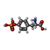
PDB-2cjz:
crystal structure of the c472s mutant of human protein tyrosine phosphatase ptpn5 (step, striatum enriched phosphatase) in complex with phosphotyrosine
Method: X-RAY DIFFRACTION / Resolution: 1.7 Å

PDB-2gjt:
Crystal structure of the human receptor phosphatase PTPRO
Method: X-RAY DIFFRACTION / Resolution: 2.15 Å

PDB-2h4v:
Crystal Structure of the Human Tyrosine Receptor Phosphatase Gamma
Method: X-RAY DIFFRACTION / Resolution: 1.55 Å

PDB-2i75:
Crystal Structure of Human Protein Tyrosine Phosphatase N4 (PTPN4)
Method: X-RAY DIFFRACTION / Resolution: 2.45 Å

PDB-2jjd:
Protein Tyrosine Phosphatase, Receptor Type, E isoform
Method: X-RAY DIFFRACTION / Resolution: 3.2 Å

PDB-2nlk:
Crystal structure of D1 and D2 catalytic domains of human Protein Tyrosine Phosphatase Gamma (D1+D2 PTPRG)
Method: X-RAY DIFFRACTION / Resolution: 2.4 Å

PDB-2nz6:
Crystal structure of the PTPRJ inactivating mutant C1239S
Method: X-RAY DIFFRACTION / Resolution: 2.3 Å

PDB-2oc3:
Crystal Structure of the Catalytic Domain of Human Protein Tyrosine Phosphatase non-receptor Type 18
Method: X-RAY DIFFRACTION / Resolution: 1.5 Å

PDB-2ooq:
Crystal Structure of the Human Receptor Phosphatase PTPRT
Method: X-RAY DIFFRACTION / Resolution: 1.8 Å

PDB-2p6x:
Crystal structure of human tyrosine phosphatase PTPN22
Method: X-RAY DIFFRACTION / Resolution: 1.9 Å

PDB-2pa5:
Crystal structure of human protein tyrosine phosphatase PTPN9
Method: X-RAY DIFFRACTION / Resolution: 1.6 Å

PDB-2qep:
Crystal structure of the D1 domain of PTPRN2 (IA2beta)
Method: X-RAY DIFFRACTION / Resolution: 2.5 Å

PDB-3b7o:
Crystal structure of the human tyrosine phosphatase SHP2 (PTPN11) with an accessible active site
Method: X-RAY DIFFRACTION / Resolution: 1.6 Å

Chemicals

ChemComp-CL:
Unknown entry / Chloride

ChemComp-NA:
Unknown entry

ChemComp-EDO:
1,2-ETHANEDIOL / Ethylene glycol

ChemComp-HOH:
WATER / Water

ChemComp-NI:
NICKEL (II) ION / Nickel

ChemComp-PTR:
O-PHOSPHOTYROSINE

ChemComp-FLC:
CITRATE ANION / Citric acid

ChemComp-ACT:
ACETATE ION / Acetate

ChemComp-SO4:
SULFATE ION / Sulfate

ChemComp-PO4:
PHOSPHATE ION / Phosphate

ChemComp-B3P:
2-[3-(2-HYDROXY-1,1-DIHYDROXYMETHYL-ETHYLAMINO)-PROPYLAMINO]-2-HYDROXYMETHYL-PROPANE-1,3-DIOL / pH buffer*YM / Bis-tris propane

ChemComp-SCN:
THIOCYANATE ION / Thiocyanate

ChemComp-MLT:
D-MALATE / Malic acid

Source
  • homo sapiens (human)
KeywordsHYDROLASE / Tyrosine Receptor Phosphatase / Beta / Human / Structural Genomics / Structural Genomics Consortium / SGC / Protein Tyrosine Phosphatase / non-receptor Type / RECEPTOR TYPE TYROSINE PHOSPHATASE J / PTPRJ / GLYCOPROTEIN / PROTEIN PHOSPHATASE / STEP / PTPN5 / PHOSPHATASE / Tyrosine phosphatase / PTPRO / GLEPP1 / PTPU2 / GAMMA / PTPN4 / PTP / MEG-1 / TRANSMEMBRANE / PHOSPHOPROTEIN / CONSORTIUM / STRUCTURAL / PTPRE / MEMBRANE / GENOMICS / PTPRG / R-PTP gamma / Protein Tyrosine Phosphatase gamma / D3S1249 / HPTPG / RPTPG / PTPG / D1-D2 catalytic domains / HYDROLASE RECEPTOR TYPE TYROSINE PHOSPHATASE J / Receptor / Lymphoid phosphatase / PEP / LYP / MEG2 / PTPN9 / PTPRN2 / PTPRP / Phogrin / IA-2 beta / autoantigen / SHP2 / PTPN11 / Deafness / Disease mutation / Phosphorylation / SH2 domain

+
About Yorodumi Papers

-
News

-
Feb 9, 2022. New format data for meta-information of EMDB entries

New format data for meta-information of EMDB entries

  • Version 3 of the EMDB header file is now the official format.
  • The previous official version 1.9 will be removed from the archive.

Related info.:EMDB header

External links:wwPDB to switch to version 3 of the EMDB data model

-
Aug 12, 2020. Covid-19 info

Covid-19 info

URL: https://pdbj.org/emnavi/covid19.php

New page: Covid-19 featured information page in EM Navigator.

Related info.:Covid-19 info / Mar 5, 2020. Novel coronavirus structure data

+
Mar 5, 2020. Novel coronavirus structure data

Novel coronavirus structure data

Related info.:Yorodumi Speices / Aug 12, 2020. Covid-19 info

External links:COVID-19 featured content - PDBj / Molecule of the Month (242):Coronavirus Proteases

+
Jan 31, 2019. EMDB accession codes are about to change! (news from PDBe EMDB page)

EMDB accession codes are about to change! (news from PDBe EMDB page)

  • The allocation of 4 digits for EMDB accession codes will soon come to an end. Whilst these codes will remain in use, new EMDB accession codes will include an additional digit and will expand incrementally as the available range of codes is exhausted. The current 4-digit format prefixed with “EMD-” (i.e. EMD-XXXX) will advance to a 5-digit format (i.e. EMD-XXXXX), and so on. It is currently estimated that the 4-digit codes will be depleted around Spring 2019, at which point the 5-digit format will come into force.
  • The EM Navigator/Yorodumi systems omit the EMD- prefix.

Related info.:Q: What is EMD? / ID/Accession-code notation in Yorodumi/EM Navigator

External links:EMDB Accession Codes are Changing Soon! / Contact to PDBj

+
Jul 12, 2017. Major update of PDB

Major update of PDB

  • wwPDB released updated PDB data conforming to the new PDBx/mmCIF dictionary.
  • This is a major update changing the version number from 4 to 5, and with Remediation, in which all the entries are updated.
  • In this update, many items about electron microscopy experimental information are reorganized (e.g. em_software).
  • Now, EM Navigator and Yorodumi are based on the updated data.

External links:wwPDB Remediation / Enriched Model Files Conforming to OneDep Data Standards Now Available in the PDB FTP Archive

-
Yorodumi Papers

Database of articles cited by EMDB/PDB/SASBDB data

  • Database of articles cited by EMDB, PDB, and SASBDB entries
  • Using PubMed data

Related info.:EMDB / PDB / SASBDB / Yorodumi / EMN Papers / Changes in new EM Navigator and Yorodumi

Read more