 ムービー
ムービー コントローラー
 構造ビューア
 EMN検索について


-検索条件
-検索結果
検索 (著者・登録者: wagner & m)の結果187件中、1から50件目までを表示しています

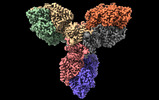
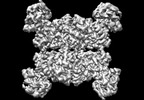
EMDB-16986: 
Structure of the relaxed thin filament from FIB milled left ventricular mouse myofibrils (tropomyosin masked out)

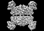
EMDB-16987:
Structure of the relaxed thin filament from FIB milled left ventricular mouse myofibrils (including tropomyosin)

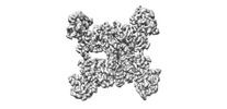
EMDB-16988:
Tomogram of sarcomere C-zone from mouse cardiac muscle

EMDB-16989:
Tomogram of sarcomere M-band to C-zone from mouse cardiac muscle

EMDB-16990:
Structure of the relaxed thick filament from FIB milled left ventricular mouse myofibrils - Crowns P2-A1

EMDB-16991:
Structure of the relaxed thick filament from FIB milled left ventricular mouse myofibrils - M-band

EMDB-16992:
Structure of the relaxed thick filament from FIB milled left ventricular mouse myofibrils - Crowns A15-A29

EMDB-16993:
Structure of the relaxed thick filament from FIB milled left ventricular mouse myofibrils - Crown P1

EMDB-16994:
Structure of the relaxed thick filament from FIB milled left ventricular mouse myofibrils - Crowns A11-A15

EMDB-16995:
Structure of the relaxed thick filament from FIB milled left ventricular mouse myofibrils - Crowns A8-A12

EMDB-16996:
Structure of the relaxed thick filament from FIB milled left ventricular mouse myofibrils - Crowns A5-A7

EMDB-16997:
Structure of the relaxed thick filament from FIB milled left ventricular mouse myofibrils - Crowns A1-A5

EMDB-18146:
In situ structures from relaxed cardiac myofibrils reveal the organization of the muscle thick filament

EMDB-18200:
Thin filament consensus map from FIB milled relaxed left ventricular mouse myofibrils

EMDB-18147:
Thin filament from FIB milled relaxed left ventricular mouse myofibrils

EMDB-18198:
Helical reconstruction of the relaxed thick filament from FIB milled left ventricular mouse myofibrils
PDB-8q4g:
Thin filament from FIB milled relaxed left ventricular mouse myofibrils

PDB-8q6t:
Helical reconstruction of the relaxed thick filament from FIB milled left ventricular mouse myofibrils

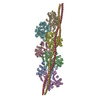
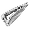
EMDB-16236:
Triangle 1 v1 for DNA ORIGAMI TRAPS FOR LARGE VIRUSES

EMDB-16237:
Triangle-1-version-2 for DNA ORIGAMI TRAPS FOR LARGE VIRUSES

EMDB-16238:
Triangle-1-version-3 for DNA ORIGAMI TRAPS FOR LARGE VIRUSES

EMDB-16239:
Triangle-2-version-2 for DNA ORIGAMI TRAPS FOR LARGE VIRUSES
EMDB-16240:
Triangle-2-version-3 for DNA ORIGAMI TRAPS FOR LARGE VIRUSES

EMDB-16261:
Triangle-2-version-1 for DNA ORIGAMI TRAPS FOR LARGE VIRUSES
EMDB-16262:
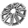
9-mer cone assembly for DNA ORIGAMI TRAPS FOR LARGE VIRUSES

EMDB-16263:
10-mer-cone for DNA ORIGAMI TRAPS FOR LARGE VIRUSES
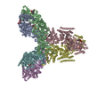
EMDB-17452:
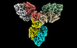
Single particle cryo-EM structure of the homohexameric 2-oxoglutarate dehydrogenase OdhA from Corynebacterium glutamicum
EMDB-17453:
Single particle cryo-EM structure of homohexameric 2-oxoglutarate dehydrogenase OdhA from Corynebacterium glutamicum with Coenzyme A bound to the E2o domain

EMDB-17454:
Single particle cryo-EM structure of homohexameric 2-oxoglutarate dehydrogenase OdhA from Corynebacterium glutamicum in complex with the product succinyl-CoA

EMDB-17455:
Single particle cryo-EM structure of homohexameric 2-oxoglutarate dehydrogenase OdhA from Corynebacterium glutamicum following reaction with the 2-oxoglutarate analogue succinyl phosphonate

EMDB-17456:
Single particle cryo-EM structure of the complex between Corynebacterium glutamicum homohexameric 2-oxoglutarate dehydrogenase OdhA and the FHA-protein inhibitor OdhI

PDB-8p5t:
Single particle cryo-EM structure of the homohexameric 2-oxoglutarate dehydrogenase OdhA from Corynebacterium glutamicum

PDB-8p5u:
Single particle cryo-EM structure of homohexameric 2-oxoglutarate dehydrogenase OdhA from Corynebacterium glutamicum with Coenzyme A bound to the E2o domain

PDB-8p5v:
Single particle cryo-EM structure of homohexameric 2-oxoglutarate dehydrogenase OdhA from Corynebacterium glutamicum in complex with the product succinyl-CoA
PDB-8p5w:
Single particle cryo-EM structure of homohexameric 2-oxoglutarate dehydrogenase OdhA from Corynebacterium glutamicum following reaction with the 2-oxoglutarate analogue succinyl phosphonate
PDB-8p5x:
Single particle cryo-EM structure of the complex between Corynebacterium glutamicum homohexameric 2-oxoglutarate dehydrogenase OdhA and the FHA-protein inhibitor OdhI

EMDB-29930:
T. cruzi topoisomerase II alpha bound to dsDNA and the covalent inhibitor CT1

PDB-8gcc:
T. cruzi topoisomerase II alpha bound to dsDNA and the covalent inhibitor CT1
EMDB-33247:
Cryo-EM structure of the Neuromedin U receptor 2 (NMUR2) in complex with G Protein and its endogeneous Peptide-Agonist NMU25

PDB-7xk8:
Cryo-EM structure of the Neuromedin U receptor 2 (NMUR2) in complex with G Protein and its endogeneous Peptide-Agonist NMU25
EMDB-27936:
Cryo-EM structure of Apo form ME3
EMDB-27937:
Cryo-EM structure of human ME3 in the presence of citrate
EMDB-27945:
Cryo-EM structure of human ME3 in the presence of citrate

PDB-8e76:
Cryo-EM structure of Apo form ME3

PDB-8e78:
Cryo-EM structure of human ME3 in the presence of citrate

PDB-8e8o:
Cryo-EM structure of human ME3 in the presence of citrate

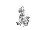
EMDB-16451:
Subtomogram average of the T. kivui 70S ribosome in situ
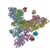
EMDB-25214:
Cryo-EM structure of Drosophila Integrator cleavage module (IntS4-IntS9-IntS11) in complex with IP6
PDB-7sn8:
Cryo-EM structure of Drosophila Integrator cleavage module (IntS4-IntS9-IntS11) in complex with IP6

EMDB-14169:
Cryo-EM structure of Hydrogen-dependent CO2 reductase.
ページ:
 wwPDBはEMDBデータモデルのバージョン3へ移行します
