 ムービー
ムービー コントローラー
 構造ビューア
 EMN検索について


-検索条件
-検索結果
検索 (著者・登録者: schmid & mf)の結果183件中、1から50件目までを表示しています
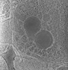
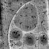
EMDB-28138: 
3D reconstruction of the apical complex of Plasmodium falciparum (3D7) free merozoite
EMDB-28141:
3D reconstruction of the apical complex of Plasmodium falciparum (3D7) free merozoite
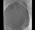
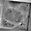
EMDB-28142:
3D Reconstruction of Plasmodium falciparum (3D7) free merozoite
EMDB-41840:
Gaussian Mixture Models based single particle refinement - GPCR (Substance P bound to active human neurokinin 1 receptor in complex with miniGs399 from EMPIAR-10786)

EMDB-41841:
Gaussian mixture model based single particle refinement - SARS (SARS-CoV-2 Spike Proteins on intact virions from EMPIAR-10492)
EMDB-41845:
Gaussian mixture model based single particle refinement - ABC transporter (inhibitor-bound ABCG2 from EMPIAR-10374)

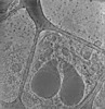
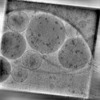
EMDB-28125:
Plasmodium falciparum merozoites apical 2-ring units

EMDB-28126:
Toxoplasma apical rings
EMDB-29207:
CryoET tomogram of mitochondria in BACHD mouse model neuron
EMDB-29208:
CryoET tomogram of BACHD mouse model neuron showing sheet aggregates
EMDB-29210:
CryoET tomogram of purified mitochondria from HD patient iPSC-derived neuron (Q109)
EMDB-29211:
CryoET tomogram of HD patient iPSC-derived neuron (Q66) with PIAS1 hetKO treatment
EMDB-28668:
CryoET tomogram of iPSC-derived control non-HD neuron (Q18)
EMDB-28944:
CryoET tomogram of iPSC-derived control non-HD neuron (Q20)
EMDB-28946:
CryoET tomogram of HD patient iPSC-derived neuron (Q53)
EMDB-29074:
CryoET tomogram of HD patient iPSC-derived neuron (Q66)
EMDB-29075:
CryoET tomogram of HD patient iPSC-derived neuron (Q77)
EMDB-29076:
CryoET tomogram of HD patient iPSC-derived neuron (Q109)
EMDB-29079:
CryoET tomogram of HD patient iPSC-derived neuron (Q66) showing sheet aggregate
EMDB-29080:
CryoET tomogram of HD patient iPSC-derived neuron (Q66) with PIAS1 hetKO treatment
EMDB-29081:
CryoET tomogram of HD patient iPSC-derived neuron (Q66) with GRSF1 KD treatment
EMDB-29083:
CryoET tomogram of mitochondria in WT mouse neuron
EMDB-29084:
CryoET tomogram of mitochondria in BACHD-dN17 mouse model neuron

EMDB-26448:
Half-bud CHIKV assembly/budding intermediate from virus-infected human cells
EMDB-25093:
Electron cryo-tomography of CHIKV assembly intermediates in human U2OS cells
EMDB-25094:
Electron cryo-tomography of CHIKV assembly intermediates in human U2OS cells
EMDB-25095:
Electron cryo-tomography of CHIKV assembly intermediates in human U2OS cells
EMDB-25096:
Electron cryo-tomography of CHIKV assembly intermediates in human U2OS cells
EMDB-25097:
Electron cryo-tomography of CHIKV assembly intermediates in human U2OS cells
EMDB-25098:
Electron cryo-tomography of CHIKV assembly intermediates in human U2OS cells
EMDB-25099:
Electron cryo-tomography of CHIKV assembly intermediates in human U2OS cells
EMDB-32904:
Structure of an orphan GPCR-G protein signaling complex

EMDB-32331:
Cryo-EM structure of the alpha2A adrenergic receptor GoA signaling complex bound to a G protein biased agonist

EMDB-32342:
Cryo-EM structure of the alpha2A adrenergic receptor GoA signaling complex bound to a biased agonist
EMDB-26447:
Docking NC CHIKV assembly/budding intermediate from virus-infected human cells
EMDB-26018:
The symmetry-released subpellicular microtubule map from detergent-extracted Toxoplasma cells
EMDB-26019:
Subpellicular microtubule from detergent-extract Toxoplasma gondii cells

EMDB-26020:
The tubulin-based conoid from detergent-extract Toxoplasma gondii cells
EMDB-26449:
Three-quarter CHIKV assembly/budding intermediate from virus-infected human cells

EMDB-26450:
Pinch-off CHIKV assembly/budding intermediate from virus-infected human cells

EMDB-26451:
Cytosolic nucleocapsid intermediate from CHIKV-infected cells
EMDB-26452:
Budding CHIKV nucleocapsid intermediate from virus-infected cells
EMDB-26446:
Electron cryo-tomography of CHIKV assembly intermediates in human U2OS cells
EMDB-31164:
Structure of the GPR88-Gi1 signaling complex bound to a synthetic ligand
EMDB-26010:
The symmetrized subpellicular microtubule map from intact Toxoplasma gondii cells
EMDB-26006:
3D reconstruction of detergent-extract Toxoplasma gondii cells at the apical end
EMDB-26007:
Three-dimensional organization of the apical complex in Toxoplasma tachyzoites
EMDB-26008:
In situ average map of conoid with PCR from intact Toxoplasma gondii cells
EMDB-26009:
The conoid segment from intact Toxoplasma gondii cells

EMDB-24665:
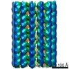
Apoferritin structure at 1.27 angstrom resolution determined from a 300 kV Titan Krios G3i electron microscope with Falcon4 detector
ページ:
 wwPDBはEMDBデータモデルのバージョン3へ移行します
